Biology Reference
In-Depth Information
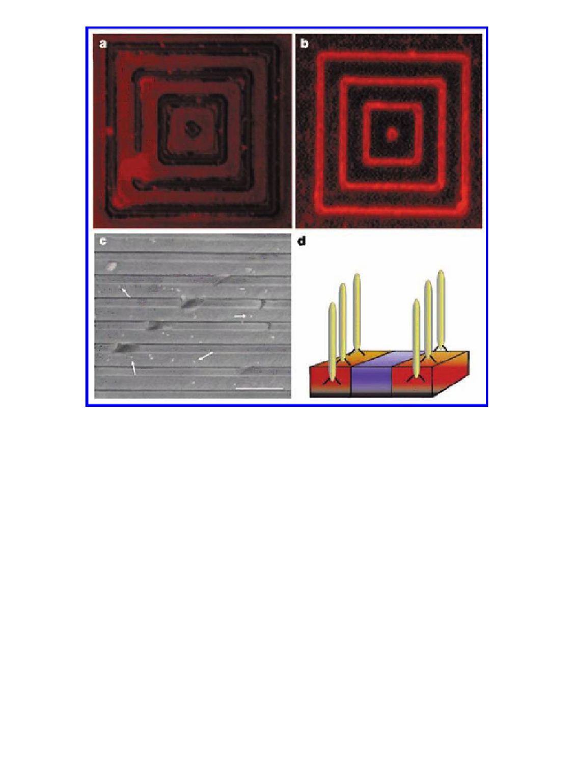
Figure
Phage recognition of semi-conductor heterostructures. (a, b)
Fluorescence images related to GaAs recognition by phage. (a) Control experiment:
no phage is present, but primary antibody and streptavidin-tetramethyl
rhodamine (TMR) are present. (b) The GaAs clone G12-3 was interacted with a
substrate patterned with 1 mm GaAs lines and 4 mm SiO
6.10
spaces. The phage was
then fluorescently labeled with TMR. The G12-3 clone specifically recognized
the GaAs and not the SiO
2
surface; scale bar, 4 mm. A diagram of this recognition
process is shown in (d), in which phage specifically attach to one semi-conductor
rather than another, in a heterostructure. (c) An SEM image of a heterostructure
containing alternating layers of GaAs and Al
2
As, used to demonstrate that
this recognition is element-specific. The cleaved surface was interacted with
the GaAs-selective G12-3 phage, and the phage was then tagged with 20 nm
gold particles. These nanoparticles (shown arrowed in (c)) are located on
GaAs and not AlGaAs layers. Scale bar, 500 nm. Reproduced with permission
from Whaley, S. R., English, D. S., Hu, E. L., Barbara, P. F., and Belcher,
A. M. (2000) Selection of peptides with semi-conductor binding specificity for
directed nanocrystal assembly,
Ga
0.98
0.02
Nature
,
405
(6787), 665-668.
Search WWH ::

Custom Search