Biology Reference
In-Depth Information
VLPs containing metallic nanoparticles or QDs are also promising
candidates for biomedical applications such as imaging and therapy. This will
be discussed in detail in Chapter 8; here one example is given to demonstrate
the feasibility of these hybrid VLPs for imaging purposes. The above-described
assembly protocol can be adapted to other viruses. Successful nanoparticle-
templated VLP assembly has been demonstrated using coat proteins from
an alphavirus (Goicochea
.,
2009), both of which are mammalian viruses. SV40 is a polyomavirus that
infects humans and monkeys. Infections are mostly latent, but the virus has
the potential for tumor transformation. SV40 is a double-stranded DNA virus.
The non-enveloped capsid has icoashedral
et al
., 2007) and
Simian virus
40 (SV40) (Li
et al
= 7 symmetry and a diameter
of 45 nm (The Universal Virus Database of the International Committee on
Taxonomy of Viruses, ICTVdB, http://www.nbci.nlm.nih.gov/ICTVdb).
Common strategies for live imaging of viral trafficking within cells utilize
bioconjugation techniques and the covalent attachment of imaging molecules
to the exterior surface of the VNP. However, when studying viral entry and
intracellular trafficking, exterior surface modifications of virus particles are
typically not desired as they can alter and interfere with the natural functions
of the capsid such as receptor recognition. Now, it has been demonstrated
that QDs with a diameter of 4 nm and coated with mercaptoacetic acid could
be encapsulated in SV40 VLPs. SV40 coat protein monomers were mixed with
the major coat protein of SV40, the VP1 protein. VLPs with
T
T =
1 symmetry
±
and a diameter of 24.1
2.2 nm were self-assembled. The utility of the hybrid
material to study viral trafficking in tissue culture has been demonstrated
(Fig. 5.11) (Li
et al
., 2009).
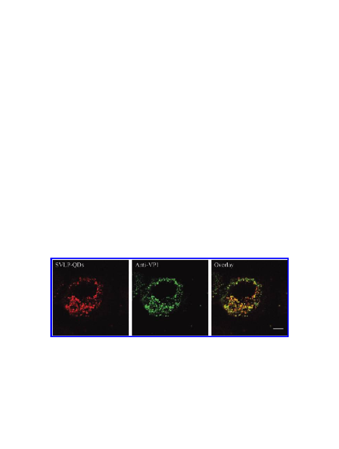
Figure 5.11
SV40-like particles with encapsulated QDs (SVLP-QDs) can ''infect'' Vero
cells. Immunofluorescence using anti-VP1 antibody in the SVLP-QD-loaded (18 h at
37.8 °C) Vero cells. The co-localization between VP1 (green) and QDs (red) confirmed
that QDs were carried into Vero cells by VLPs. Scale bars: 10
µ
m. Reproduced with
permission from Li, F., Zhang, Z. P., Peng, J., Cui, Z. Q., Pang, D. W., Li, K., Wei, H. P., Zhou,
Y. F., Wen, J. K., and Zhang X. E. (2009) Imaging viral behavior in mammalian cells
with self-assembled capsid-quantum-dot hybrid particles,
Small
,
5
(6), 718-726.

Search WWH ::

Custom Search