Biology Reference
In-Depth Information
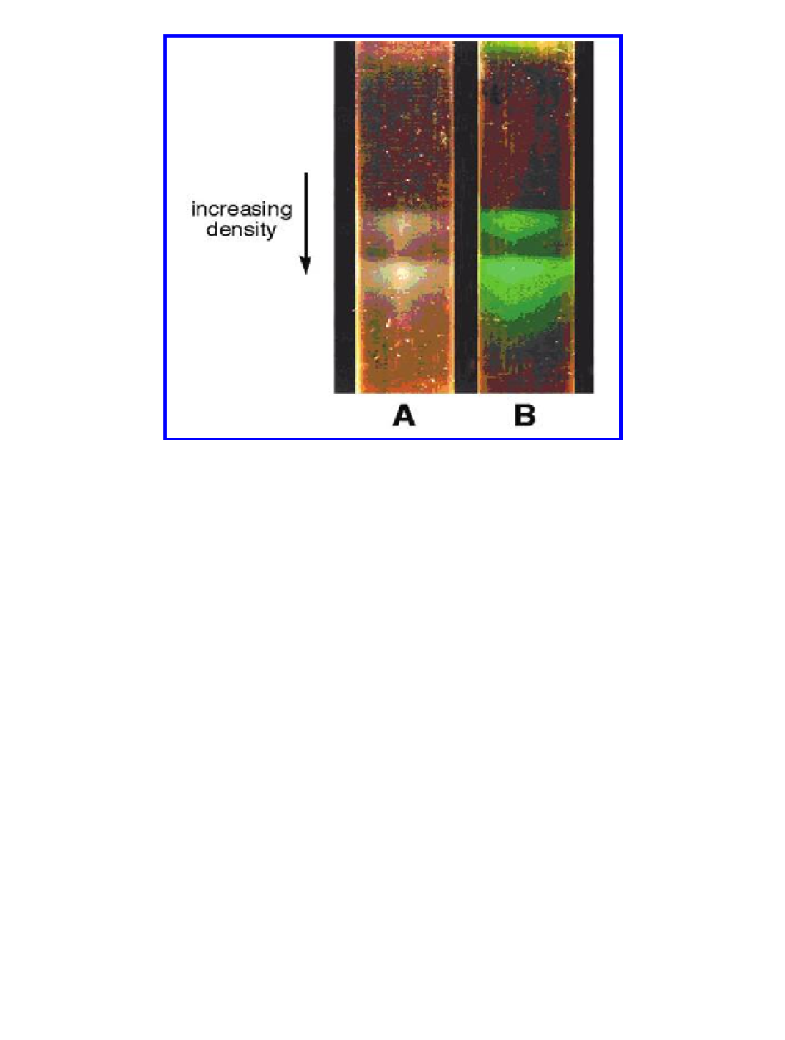
Figure 4.18
Analysis of dye-labeled VNPs. Ultracentrifugation of native (A) and
fluorescein-labeled CPMV particles (B) through sucrose gradients. The two bands in
each sample contain virus particles encapsulating the two different RNA molecules,
RNA-1 and RNA-2, of the genome and are referred to as bottom and middle
components, respectively. Top components, which are devoid of RNA, only make a
small amount of most CPMV preparations and are not visible here. Reproduced with
permission from Wang, Q., Kaltgrad, E., Lin, T., Johnson, J. E., and Finn, M. G. (2002)
Natural supramolecular building blocks: wild-type cowpea mosaic virus,
Chem. Biol.
,
9
(7), 805-811.
. APPlICAtIonS oF CheMICAlly lABeled VnPs
The VNP serves, in many cases, as a template or scaffold for functionalization.
It can be regarded as a platform for presentation of functional ligands.
VNPs comprise many copies of identical coat protein subunits and are thus
highly polyvalent. Polyvalent display is desired for a range of applications,
especially for applications in medicine (discussed in Chapter 8). Multivalent
display is also beneficial for the development of novel sensors; multivalent
display of signaling molecules leads to signal enhancement. VNPs have
been combined with redox-active molecules to yield novel electro-active
materials for potential applications in sensors, catalytic devices, or electronic
devices. Dye-labeled VNPs sensors have been utilized in biological assays for
Search WWH ::

Custom Search