Biology Reference
In-Depth Information
added mutant particles (display 180 free Cys side chains). Wild-type and
passivated mutant particles did not bind onto gold surfaces. Non-treated
Cys mutants formed extensive aggregates. Only the symmetry-broken
particles facilitated formation of a controlled monolayer on the substrate
(Fig. 4.14) (Klem
et al.
, 2003).
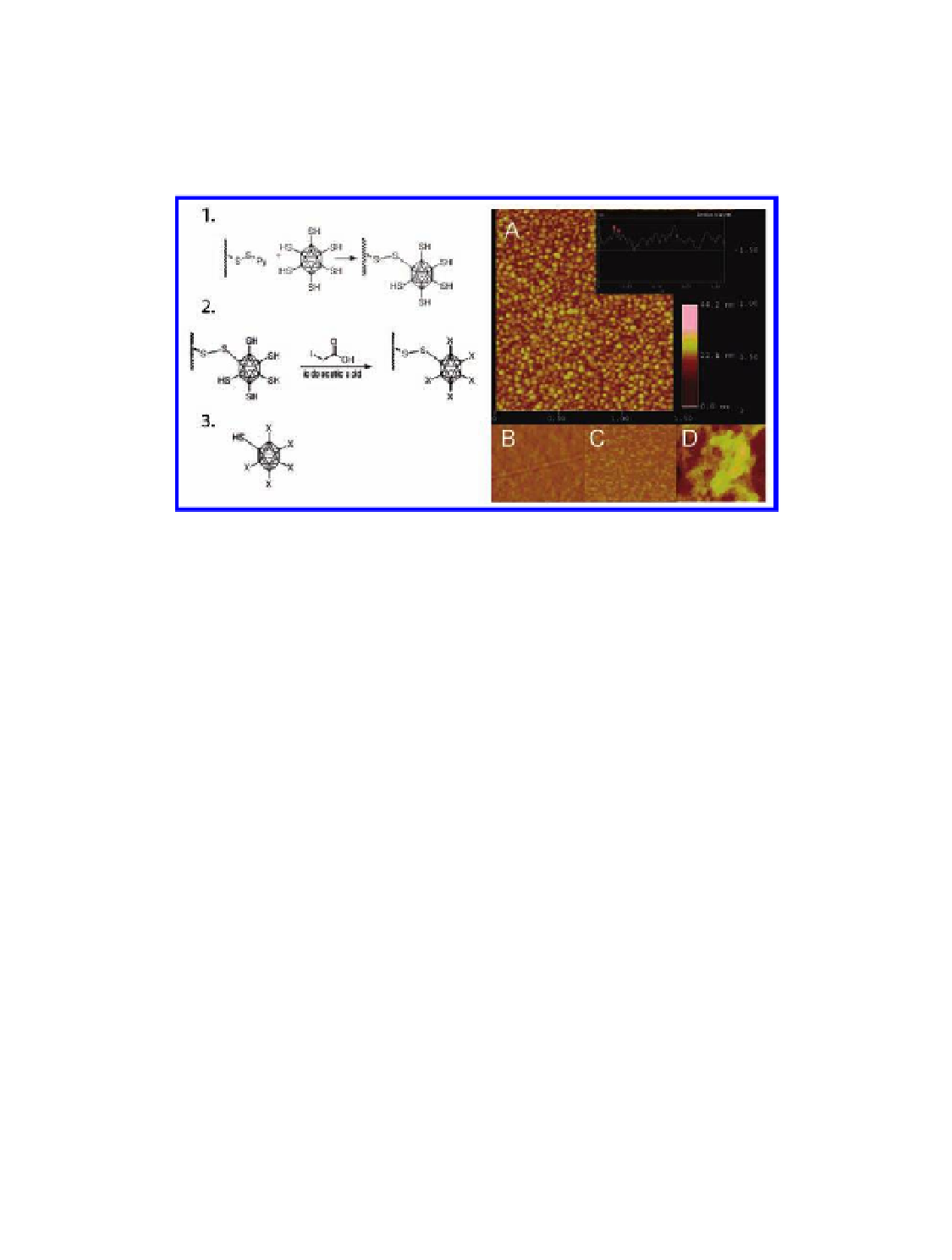
Figure 4.14
Symmetry breaking of a Cys-added mutant CCMV particle. Left panel:
outline of the solid-state syntetic approach. Step 1: binding of the particles to the
thiol-activated resin. Step 2: passivation of unbound cysteine residues with IAA. Step
3: elution of the symmetry-broken particles by reduction. Right panel: tapping-mode
atomic force microscopy imaging on CCMV particle formulations added onto a gold
substrate: (A) symmetry-broken particles with distance and height profile (inset);
(B) wild-type CCMV with no exposed thiols; (C) Cys-mutant treated with IAA to
passivate all exposed thiols; (D) untreated Cys mutant particles with 180 surface-
exposed thiols. All scans shown are 2
µ
m in length. Reproduced with permission
from Klem, M. T., Willits, D., Young, M., and Douglas, T. (2003) 2-D array formation
of genetically engineered viral cages on Au surfaces and imaging by atomic force
microscopy,
J. Am. Chem. Soc.
,
125
(36), 10806-10807.
In a different approach, breaking the symmetry was achieved by making
use of
dis- and re-assembly techniques; controlled and sequential
ligand display was facilitated through mixed self-assembly (Fig. 4.15)
(Gillitzer
in vitro
, 2006). Herein, CCMV capsids were independently decorated
with two different types of ligands to generate two populations of labeled
virions: type I labeled with ligand A (biotin) and type II labeled with ligand
B (digoxigenin). The particles were then
et al.
disassembled and the
resulting coat protein subunits separately purified. Re-assembly was
performed using controlled ratios of type I and II subunits, exerting control
of the stoichiometry of ligands A and B displayed on the final assembled
virions (Fig. 4.15) (Gillitzer
in vitro
et al.
, 2006).
Search WWH ::

Custom Search