Biology Reference
In-Depth Information
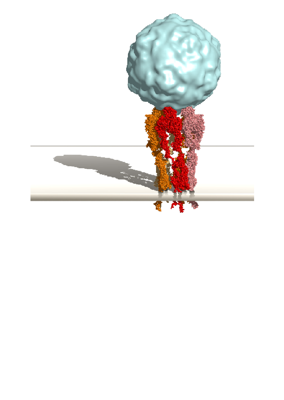
Fig. 1.
Molecular model of EV1 with three
1 integrins. EV1 is drawn
as a cyan-colored isodensity surface of a map computed at 15 Å resolution
from the high-resolution coordinates of the EV1 crystal structure (PDB
entry 1EV1).
17
α
2
β
1 integrin heterodimer in
the active conformation are drawn as space filling models in different shades
of red and orange. The model of the integrin heterodimer is based on the
crystal structures of
Three copies of a model of
α
2
β
3
(1TYE)
20
ectodomains, and published cryoelectron microscopy reconstruc-
tions. The binding site of the
α
2I domain (1AOX),
18
α
V
β
3 (1JV2)
19
and
α
IIb
β
α
2I domain on the surface has been solved
by cryo-electron microscopy.
16
I
domain and the domains of the “stalk” region within the integrin het-
erodimer were modeled by hand and eye. Three copies of the integrin het-
erodimer fit without steric hindrace into the adjacent binding sites around
the five-fold symmetry axis of the EV1 capsid. The image was made with
PyMol 0.99 (DeLano Scientific, San Carlos, CA, USA) and POV-Ray 3.6
(www.povray.org).
The orientation and position of the
α