Biology Reference
In-Depth Information
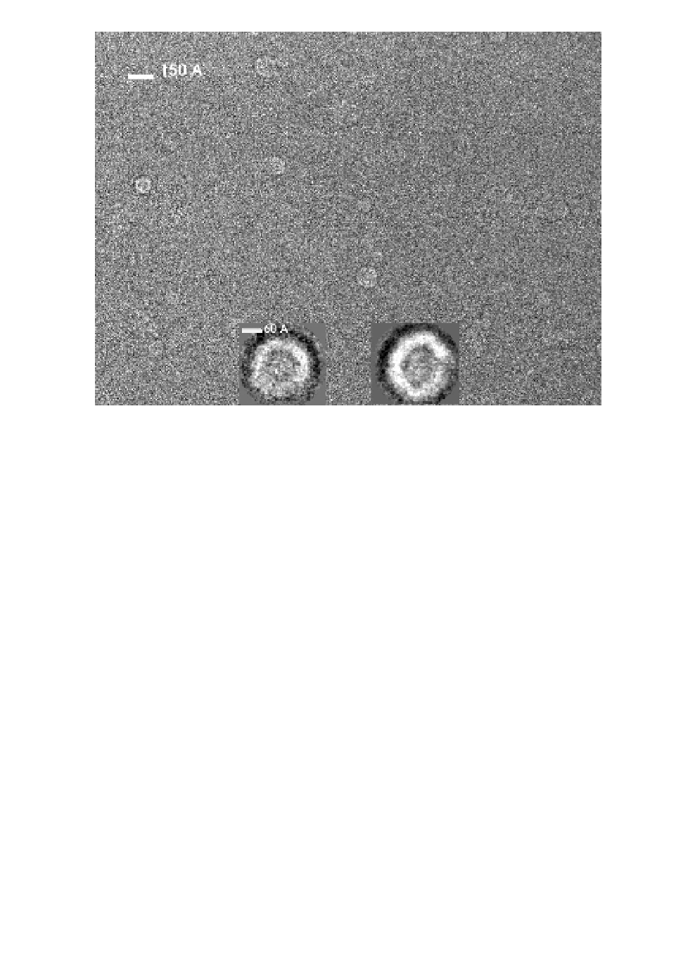
Fig. 6.
Cryo-EM preparation of purified recombinant Puumala virus
N protein (see Fig. 2). The standard cryo-EM preparative method was used
after a 3
L sample was applied to glow-discharged holey-carbon 300- or
400-mesh-Au grids (Quantifoil) washed from urea (i.e. floated in sequence in
drops of 5 mM Tris-HCl, pH 8.0). In the cryo-EM images, rings and arches
are visible that suggest higher-order packing of RNP. In the insets, class aver-
ages of particles are shown at higher magnification. Reliable 3D reconstruc-
tions were difficult to obtain due to heterogeneities in particle sizes. The
class averages were calculated using reference-free classification with EMAN.
µ
during preparation of samples. The monomers in these structures
appeared to be tightly packed, because no indications of cavities were
seen. Tight packaging of the twisted monomers in the N-protein
oligomers is in agreement with recent crystallographic data on RNPs
of rabies and vesicular stomatitis viruses.
13,14
Concluding Remarks
A single-particle reconstruction with three-fold axis of rotational sym-
metry (C3) revealed the existence, in addition to monomers, of an