Biology Reference
In-Depth Information
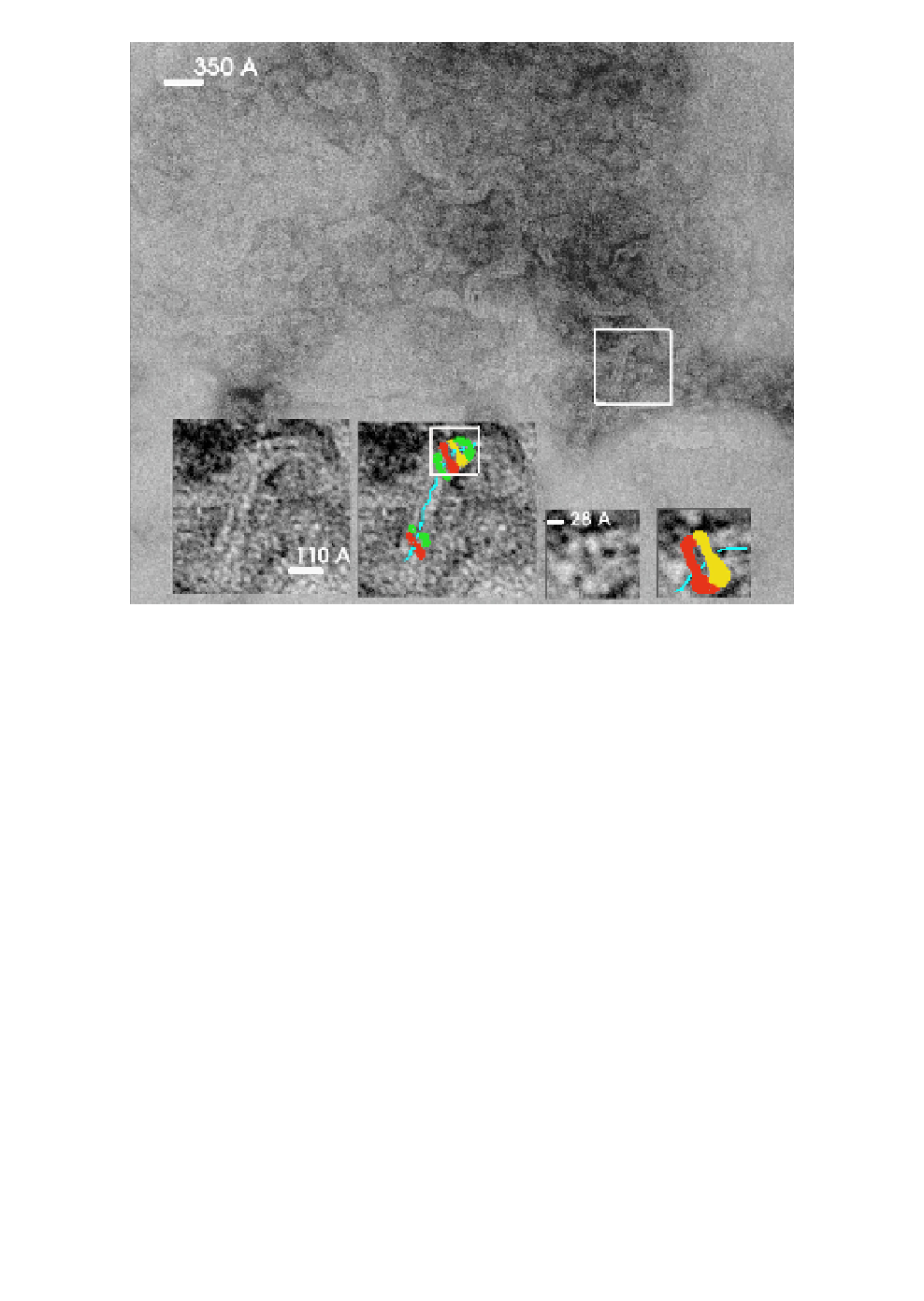
Fig. 5.
Fibrous RNP complexes formed by recombinant hantaviral N pro-
tein and cellular RNA. The fibrous complexes seen in the background are
interpreted as N protein associated with RNA. They are detected in samples
negatively stained with uranyl acetate (see Fig. 2). The selected region (framed
white) is shown at higher magnification in the two insertions at the bottom
left. The N-protein monomers are colored red, green, and yellow, and the pos-
sible track of underlying RNA that the N-monomers are covering is shown as
a blue line. The white frame in the insertion is shown in detail as a pair of
smaller insertions to the right; identified monomers of the N protein are col-
ored red and yellow and the suggested track of RNA is shown in blue. Our
interpretation of the arrangement of N-monomers in fibrous complexes is in
remarkable agreement with the models presented for the rabies virus nucleo-
capsid (cf. Figs. 6b and 6d in Schoehn
et al.
, 2001
29
).
in size and shape, to structures formed by recombinant N proteins of
influenza
28
and rabies
29
viruses and, most probably, are oligomers of
the N protein with cellular RNA wrapped. The rings of this size can
accommodate 10 to 12 monomers of the hantaviral N protein; they
are probably produced from larger structures by breaking the RNA