Biology Reference
In-Depth Information
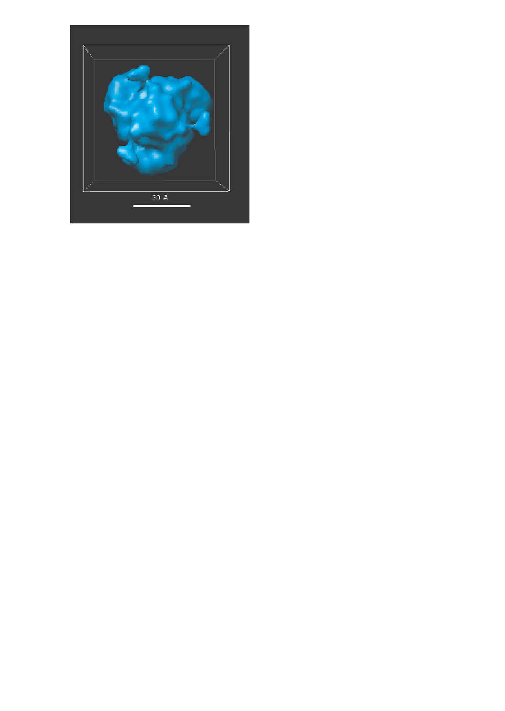
(a)
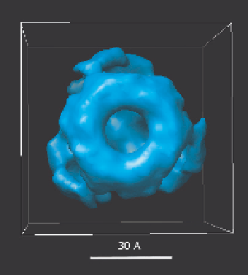
(b)
(c)
Fig. 2.
3D reconstruction of the hantaviral N-protein trimer performed
using a single particle reconstruction. The reconstruction was done assum-
ing symmetry (C3), i.e. single three-fold axis of rotational symmetry. 3
L
sample of purified recombinant Puumala virus N protein
26
(0.5-1 mg/mL,
dissolved in 6 M urea, 10 mM Tris-HCl, pH 8.0) was applied to carbon
film-coated 300- or 400-mesh-Au grids (Quantifoil), diluted and washed,
i.e. floated in sequence in drops of 1% uranyl acetate and negatively stained.
Electron microscopic pictures were collected at magnification of 50,000
at 80 kV with a Jeol 1200EX microscope. Negatives were scanned at 4,000
µ