Biology Reference
In-Depth Information
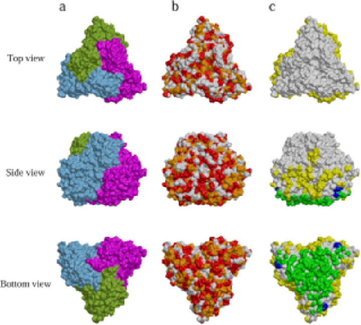
Fig. 7.
Distribution of variations in the sequence of P8 between RDV and
RGDV.
21
Surface representations are shown of P8-trimers viewed from the
top, side and bottom.
(a)
The three P8 subunits in a P8-trimer are colored blue,
magenta and green.
(b)
Regions of identical sequence in P8 of RDV and RGDV
are colored red, regions of similar sequence are colored orange, and regions of
non-similar sequence are colored white.
(c)
The regions that make contact with
neighboring P8-trimers and the P3 layer are highlighted. The surfaces in
contact with neighboring P8-trimers, with the P3 layer, and with both neigh-
boring P8-trimers and the P3 layer are colored yellow, green and blue, respec-
tively. Representations were produced with DINO (http://www.dino3d.org).
serologically or biochemically. Three-dimensional homology map-
ping of P8 proteins in RDV and RGDV (Fig. 7) indicated that the
amino acid residues at the interface between the T-trimer and the core
particle — where heterologous P3 core and P8 outer capsid proteins
interact — were more strongly conserved than those that on the upper