Biology Reference
In-Depth Information
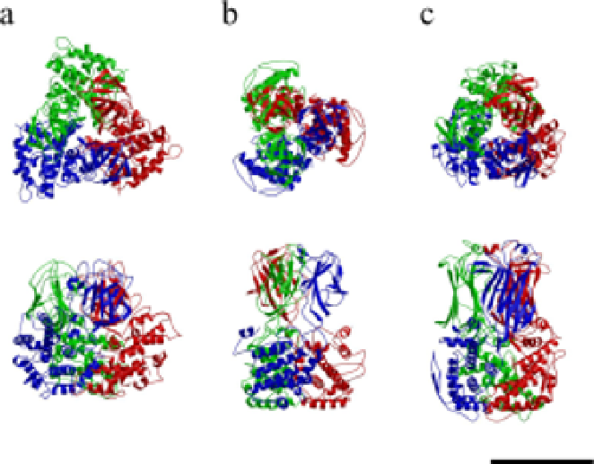
Fig. 2.
Comparison of P8 of RDV with VP7 of BTV and VP6 of rotavirus.
Top: Trimers of P8 of RDV
(a)
, VP7 of BTV
(b)
, and VP6 of rotavirus
(c)
,
as observed from the outside of the viral particle. Individual subunits are
colored red, blue, and green, respectively. Bottom: The same images after
rotation through 90
°
.
that allow the bilateral binding of homologous P3 proteins that
results in the self-assembly of a spherical particle.
When our group analyzed the structure of P3 by X-ray crystallog-
raphy,
2
we found that subunits A1 and B1 and their equivalent pairs (see
Fig. 1(a)) form the most abundant hydrogen bonds and salt bridges
among the possible P3-P3 interfaces. The amino-terminal arm of 30
residues of P3B interacts intimately with P3A (Fig. 4), and this arm
seems to stabilize the dimer of the two P3 subunits. Such an atomic
structure probably corresponds to the dimers of P3 protein, whose for-
mation was demonstrated biochemically.
8
The formation of the dimeric
structure — which can be considered to be the unit piece in the jigsaw
puzzle that yields the icosahedral core structure when completed —
might initiate the structural assembly of the RDV core particle.