Biology Reference
In-Depth Information
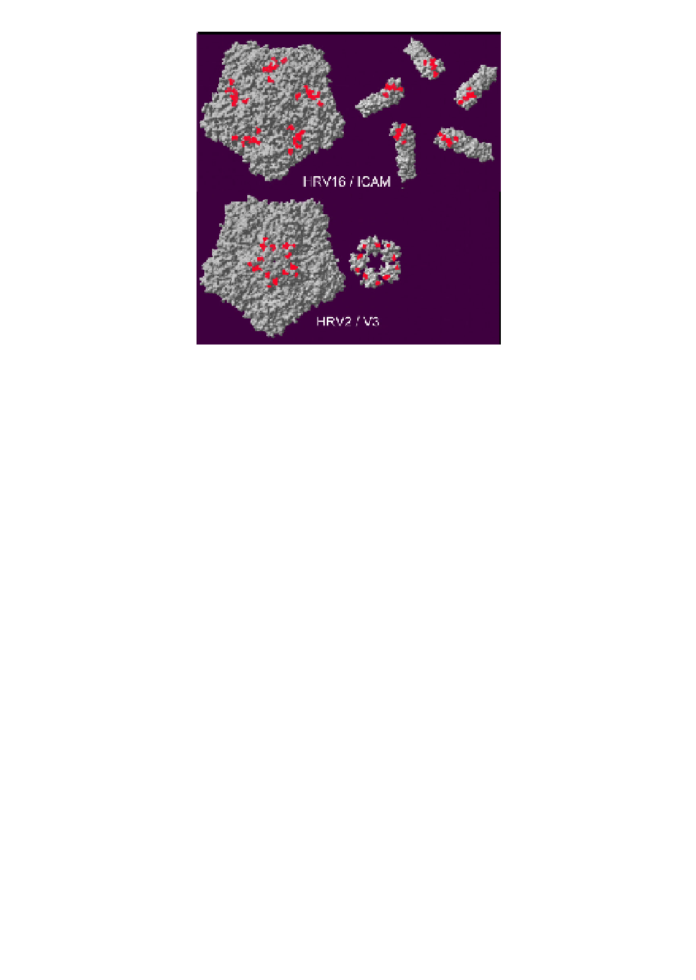
Fig. 3.
Comparison of the binding sites of V3 and of ICAM-1 on the
respective surfaces of HRV2 and HRV16. View onto a viral pentamer. The
receptors were tilted by 180
to the right to visualize the respective contacts.
Residues in HRVs and in the receptors within a distance of
°
3 Å from each
other are colored red. Note that the five individual molecules of V3 come so
close to each other that they appear to form a ring.
≤
(Fig. 3). This could either result from the binding of five copies of V3
to the five symmetry-related sites, with each V2 moving freely, or from
simultaneous binding of V2 and V3, contributed from the same mole-
cule, to two neighboring symmetry-related sites, leaving one site free.
This should give rise to a stoichiometry of 48 modules per virion (i.e.
an occupancy of 80%), as was indeed found in the reconstructions.
58
Although the amino acid sequences of V2 and V3 differ in quite a
number of positions, except those in contact with the virus that are at
least functionally conserved, the fitting into the electron density was
ambiguous; cryo-EM and X-ray crystallography are averaging tech-
niques that do not easily allow differentiation between these possibili-
ties. Therefore, under the conditions of crystal formation either 80%
of the binding sites on the virus were occupied by V3 modules only,
or each vertex was occupied by two copies of the concatemer V23.