Biology Reference
In-Depth Information
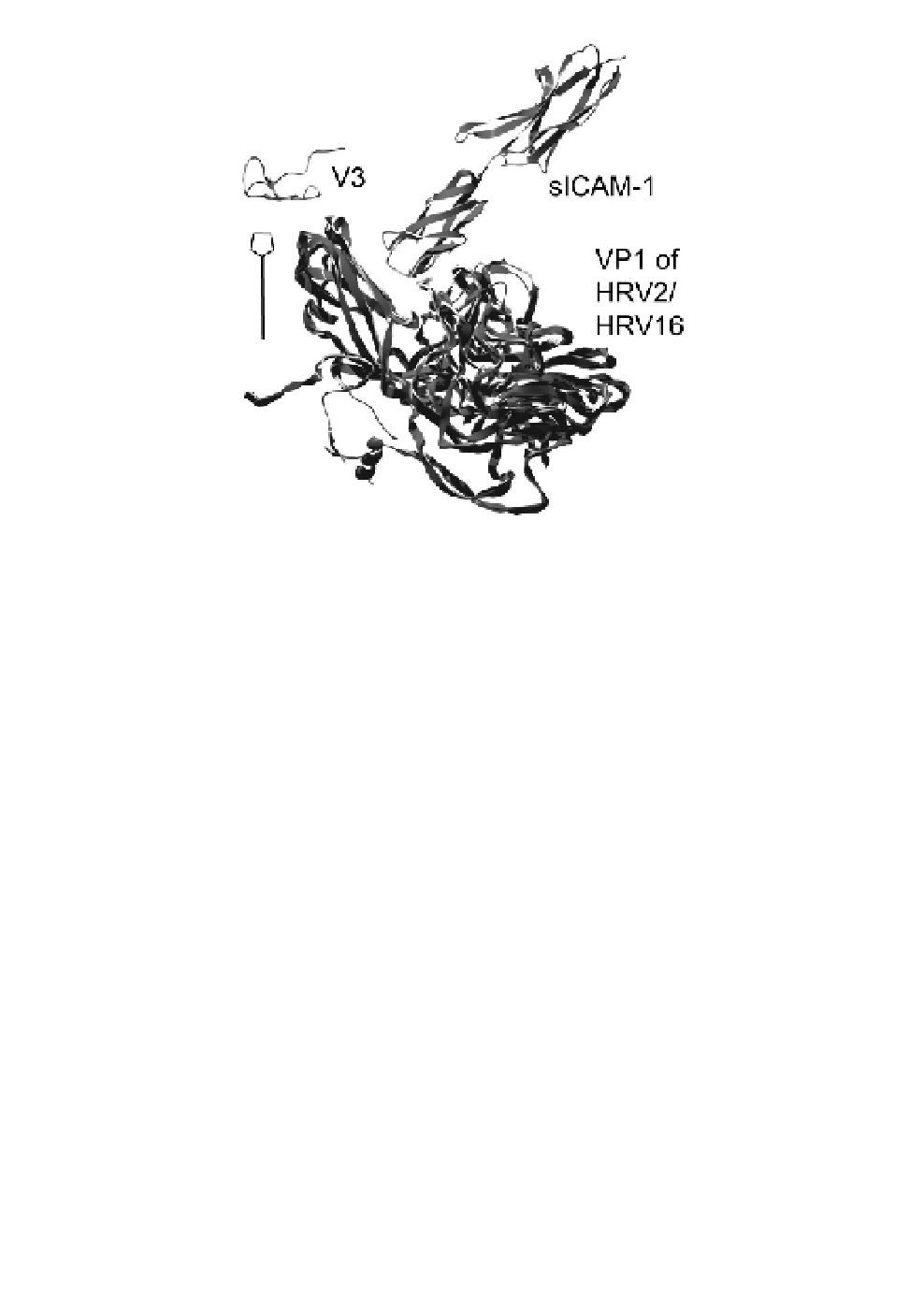
Fig. 2.
Arrangement of V3 and ICAM-1 with respect to their cognate HRVs
as seen in the complexes solved by cryo-EM (HRV16; PDB entry 1d3e)
and X-ray (HRV2; PDB entry 1v9u). Side view onto a slightly inclined VP1 of
HRV2 and HRV16 with the respective soluble recombinant receptor
fragments attached. Coordinates of VP1 with attached receptor fragments
from the cryo-EM structures of the complex between HRV16 and a two-
domain ICAM-1 (PDB entry 1d3e) and of the X-ray structure of the com-
plex between HRV2 and V23 (PDB entry 1v9u) were superimposed. The
approximate position of the five-fold axis of icosahedral symmetry is indicated.
be in contact with the canyon, whereas the second domain sticks out
into the solvent. In the cryo-EM structure of a complex between
Coxsackievirus 21 (Cox21), an enterovirus also binding ICAM-1,
and the entire exodomain of ICAM-1, all the five Ig-domains are vis-
ible, although the density of domains four and five is very low.
59
This
indicates that ICAM-1 is extremely rigid. The model also gives an
impression of the distance between the virus and the plasma mem-
brane when attached to the host cell.
59
In contrast, in the X-ray
structure of the complex between V23 and HRV2, the receptors are
arranged around the five-fold axis resulting in a ring-like appearance