Biology Reference
In-Depth Information
high-resolution cryo-EM images,
20
and even thin section EM images
22
provide a rather smooth, featureless topographical appearance for the
virion. In contrast, high-resolution
in situ
AFM images demonstrate
a high density of protrusions on the virion surface, giving it a “knobby”
appearance. The protrusions appear to be of a fairly uniform size of
about 25
30 nm, though irregular in their arrangement. In some of
the most detailed images (Fig. 5a), subunits of ~6 nm diameter can be
discriminated within the protrusions. This would correspond to a
molecular weight of about 120-150 kDa for a monomeric globular
protein. Since the molecular weights of the 16 major membrane
×
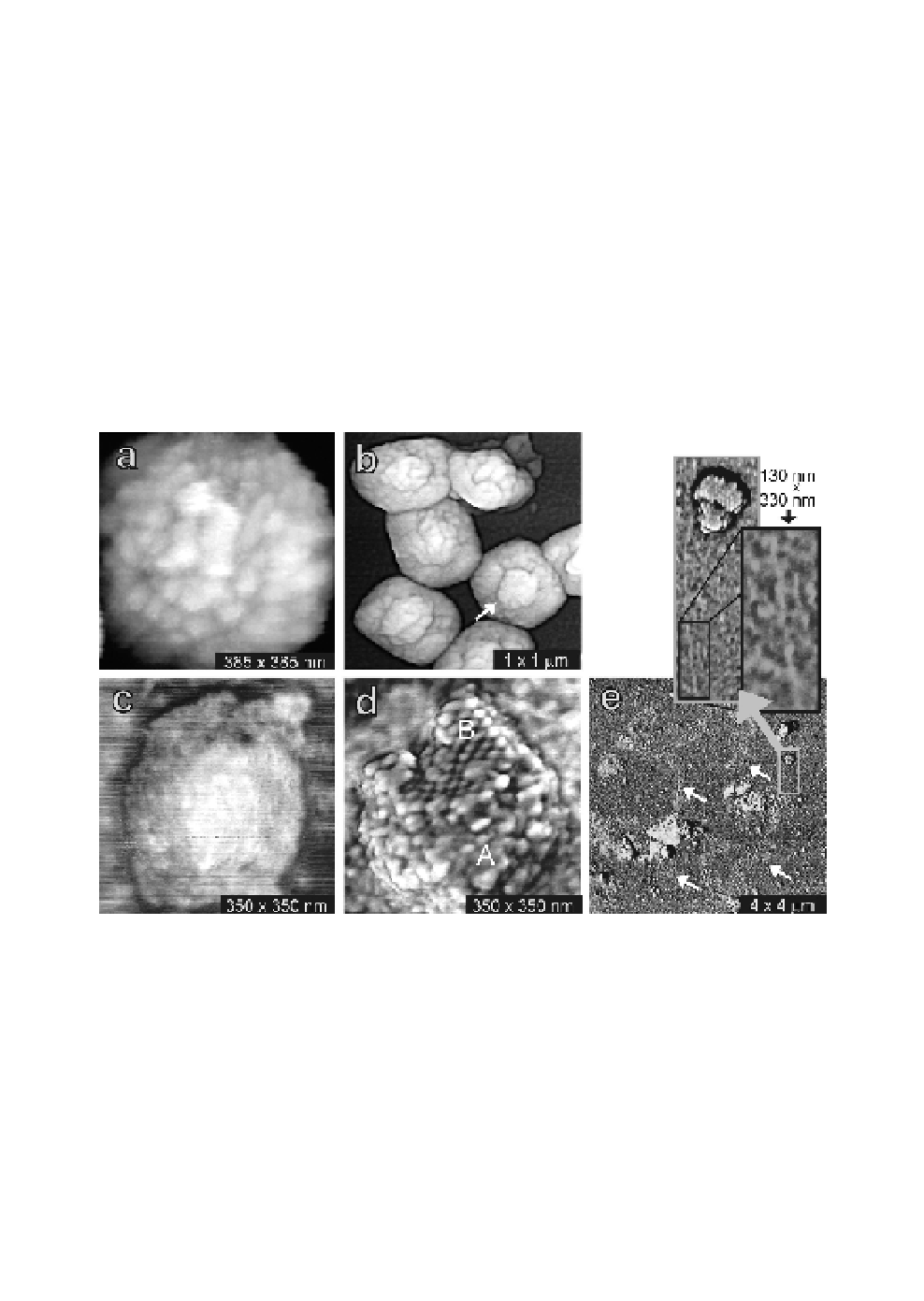
Fig. 5.
(
a
) High-resolution image of fully hydrated IMV virion in buffer.
(
b
) Air-dried IMV virions show a central raised area (arrow). (
c
) HSV-1
virion still covered in its lipid envelope. (
d
) Addition of 0.2% Triton X-100
results in removal of the envelope, showing tegument proteins (A) and the
capsid with individual capsomeres (B). (
e
) DNA escapes from HSV-1 virions
after treatment with 0.5% SDS (white arrows). The area in the grey rectangle
is shown in the two insets at higher resolution. Here, an individual double-
stranded DNA string is seen to escape a damaged virion.