Biomedical Engineering Reference
In-Depth Information
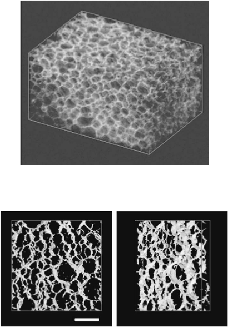
Figure 8.
FIB-SEM image of DPNR-graft-PS with the nanomatrix structure before annealing. After
aligning the observed surfaces sliced with the focused ion beam at each 102 nm interval, a three-
dimensional image in micro-metric scale was made for the rubber with nanomatrix structure. Box
size of the three-dimensional image is 6045 nm, 4123 nm and 5000 nm in X, Y, and Z directions,
respectively.
Figure 9.
Three-dimensional TEMT image. To focus on the nanomatrix, the film was stained with
RuO
4
and the matrix was colored in the image; hence, transparent domains represent natural rubber
and light yellow domains represent polystyrene. A mirror image indicates a three-dimensional
picture. Scale bar shows 1 mm. Box size of the three-dimensional image is 3740 nm, 3740 nm, and
472 nm in X, Y, and Z directions, respectively.

Search WWH ::

Custom Search