Biomedical Engineering Reference
In-Depth Information
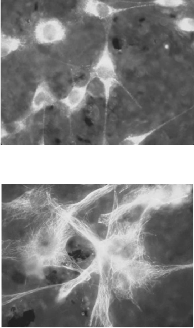
Figure 3.
MC3T3 cells on the active area of the device immediately after mechanical stimulation.
Indirect immunofluorescence using primary antibody against actin (Laminin, Ab-1, Thermo
Scientific, used at 1:50) and secondary antibody (Chromeo™ 488 conjugated Goat anti-Rabbit IgG,
Active Motif 1:500); (400X, microscope Olympus BX41, Olympus Cell A Imaging Software).
Figure 4.
MC3T3 cells on the active area of the device immediately after mechanical stimulation.
Indirect immunofluorescence using primary antibody against actin (Tubulin
b
, Thermo Scientific,
used at 1:50) and secondary antibody (Chromeo™ 488 conjugated Goat anti-Rabbit IgG, Active
Motif 1:500); (400X, microscope Olympus BX41, Olympus Cell A Imaging Software).

Search WWH ::

Custom Search