Biomedical Engineering Reference
In-Depth Information
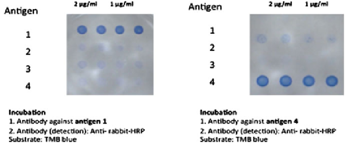
Fig. 9
Example of a 4plex miniaturised ELISA with colorimetric detection
sulfuric or phosphoric acid with maximum absorbance at 450 nm. TMB is very
sensitive and may produce significant background signal if too much protein or
antibody is used. TMB is more quickly oxidized than other HRP substrates,
resulting in faster colour development.
The development of the experimental conditions for colorimetric detection in
multiplex assays using multiple analytes in a single well as capturing agents
typically needs several rounds of optimisation, especially when the agents show
potential cross reactivity. Very essential parameters do include the actual
concentration of the printed analyte, the buffer composition, the spotting volume,
blocking reagents and times as well as timings for the individual incubation steps
and their respective reagents concentration. The development of a multiplexed
panel with TMB readout is illustrated in Fig.
9
. This figure shows the printing
of four different antigens in two different concentrations onto the bottom of a
sciPLEXPLATE. Each concentration is deposited in neighbouring duplicates.
After incubation and washing of two different antibodies in two separate wells
detection of the specific interactions was easily visible although some background
in the non specific binding spots is visible. Using an integration of the spots signals
and the introduction of a threshold clearly distinguishes specific from unspecific
binding (data not shown) .
Typically colorimetric detection is thought to be single colour detection only,
but recent developments have shown that a dual colour detection scheme is com-
patible with microplate based read-outs. In Fig.
10
different colour TMB substrates
developed by Seramun (Heidesee, Germany) were applied in a dilution series and
gave good signal to noise ratios. Two different arrays in two different wells of a 96
well sciPLEXPLATE were printed with various volumes of capturing agents. From
left to right the spotting volumes in each row are 1-2- 3 and 4 nL. While incubation
in the left well was finally done with TMB blue, in the right well a similar chemical
called TMB green was applied resulting in a green colour for detection .