Biology Reference
In-Depth Information
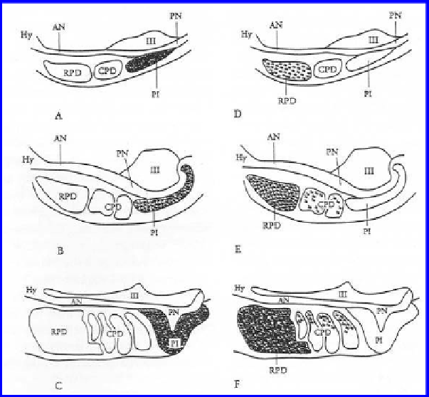
Figure 16.
Diagrammatic representation of proopiomelanotropin (POM; A, B, C) and
proopiocortin (POC; D, E, F) expression in the adenohypophysis of larval (A, D), stage 5 (B,
E) and upstream migrant (C, F)
Petromyzon
marinus
. Spatial and temporal expression patterns
were determined using
in situ
hybridization and riboprobes specifi c to POM and POC mRNAs,
respectively. Shading is indicative of relative expression levels. POM expression is restricted to
the pars intermedia (PI) throughout development. POC is expressed in the in the rostal pars
distalis (RPD) throughout development; in addition, POC expression in scattered throughout
the caudal pars distalis (CPD) during metamorphosis and in the dorsal aspect of the CPD
in the upstream migrant. Anterior neurohypophysis, AN; hypothalamus, Hy; posterior
neurophypophysis, PN; third ventricle of the brain, III. (From Ficele et al
.
, 1998).
of the RPD prevented metamorphosis and removal of the CPD resulted in
metamorphic stasis at stage 3 (Joss, 1985). The increase in POC expression
in the RPD during metamorphosis and its appearance in the CPD at stage
5 further support these fi ndings, as does the observation that ACTH
immunoreactivity increases in these cells at these stages of development
(Nozaki et al
.
, 2008). Collectively, these fi ndings suggest that ACTH, a major
POC product, may be important in lamprey metamorphosis as has been
suggested for amphibians.
POM expression was detected in most cells of the PI throughout all life
cycle stages (Fig. 16). POM signal density in immediately premetamorphic
animals was signifi cantly greater than in all stages examined except spawning
adults. Signal density decreased during stages 1-5 of metamorphosis, then