Biology Reference
In-Depth Information
of the cranial principal islet in
P. marinus
, a major contribution is through
budding and profi leration of cell clusters from the epithelium of the
intestinal diverticulum (Elliott and Youson, 1993a, 1993b). The caudal
principal islet in
P. marinus,
and likely most other northern hemisphere
species (Youson
et al
.
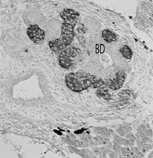
, 1988; Youson and Elliott, 1989), develops from a
transformation/dedifferentiation and proliferation of the extrahepatic,
and some intrahepatic, epithelial cells of the larval common bile duct (Fig.
14). Insulin (B) cells appear fi rst and then eventually are accompanied by
somatostatin-containing (D) cells (Elliott and Youson, 1987,1993b). The
development of the caudal principal islet is highly synchronized and
any deviation can alter the normal distribution of pancreatic islets that
make up this principal islet (Youson and Cheung, 1990). The absence of a
caudal principal islet in southern hemisphere species is due to the
complete regression of the larval bile duct epithelium, seemingly because it
enters the cephalic end of an intestinal diverticulum (Hilliard et al., 1985;
Youson, 2000).
Figure 14.
Light micrograph showing the caudal principal islet developing from the bile duct
epithelium (BD) during stage 4 of metamorphosis in
P. marinus
. Immunohistochemical staining
shows positive brown staining of islet tissue with an insulin antiserum. X270.