Biology Reference
In-Depth Information
the expression of THRβs increases in climax, reaches its peak in post
climax and remains high in metamorphosed juveniles (Fig. 15).
In situ
hybridization revealed the ubiquitous expression of THR genes and distinct
tissue specifi city of α and β subtypes in the fi sh body: THRα is strongly
expressed in tissues such as the skeletal muscle (Fig. 16) and epithelial
and immature glandular cells of the stomach (Fig. 17), which undergo
marked change (see gastric development and muscle development of this
chapter), while THRβ is strongly expressed in tissues that do not show
apparent changes. Collectively, these results indicate the possibility that,
unlike amphibians, αtype (αA) THR rather than βtype may play important
roles in metamorphic changes in the Japanese fl ounder. These results also
strongly suggest that the development of tissues stimulated by thyroid
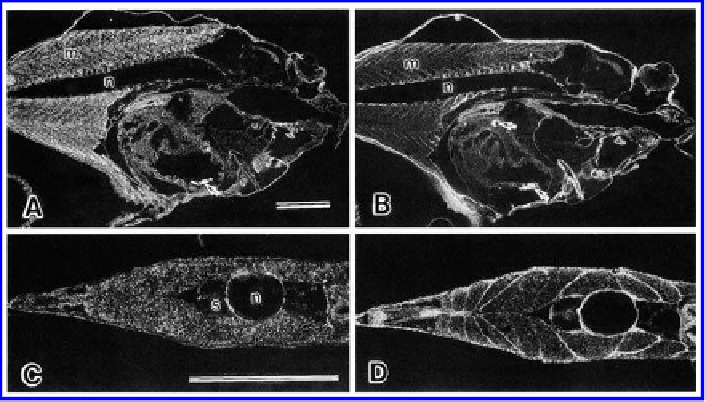
Figure 16.
Dark-fi eld photomicrographs of the detection of TRα (A and C) and TRβ (B and
D) mRNAs in two adjacent sagittal (A and B) and transverse (C and D) sections of Japanese
fl ounder larvae at metamorphic climax. Arrows indicate nonspecifi c signal from ingested
food, m, skeletal muscle; n, notochord; s, spinal cord. Bar=1mm. [From Yamano and Miwa,
1998, with permission].
hormone during metamorphosis is further enhanced and/or controlled
at the receptor level by stage- and region-specifi c expressions of THRs
(Yamano and Miwa, 1998).
4.2.7 Modifi cation of thyroid hormone actions by other hormones
4.2.7.1 Cortisol
In anuran amphibians, adrenal corticosteroids are known to enhance effects
of thyroid hormones in metamorphosing tadpoles (Frieden and Naile, 1955;