Biomedical Engineering Reference
In-Depth Information
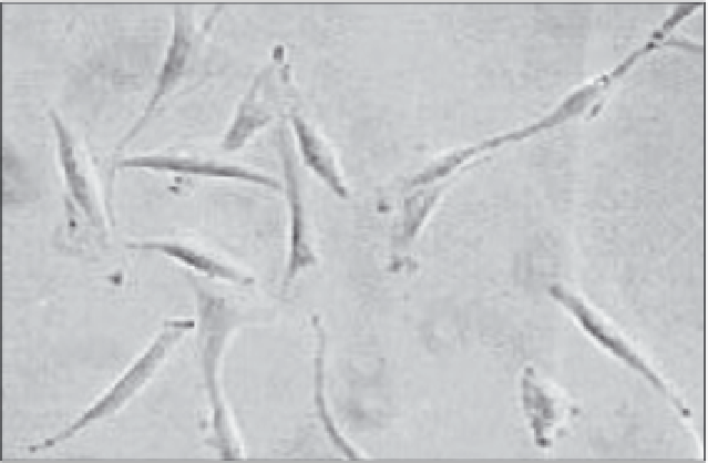
Figure 1. Phenotype of cultured human BMSC. BMSC cultured in the presence of FGF-2 maintain a stable
fibroblastic phenotype.
purpose indeed a number of factors has to be considered: minimal invasivity, miminal donor
site morbidity, easily accessible tissue source, high frequency of osteoprogenitors etc.
Marrow derived stromal fibroblasts (BMSC) (Fig. 1) can be isolated, expanded in culture,
and stimulated to differentiate into bone, cartilage, muscle, marrow stroma, tendon, fat and a
variety of other connective tissues
41
(Fig. 2). The first evidence for BMSC differentiation po-
tential derives from transplantation studies in animal models performed by Friedenstein and
coworkers.
39
The harvest of a limited bone marrow sample is a relatively easy and safe proce-
dure. Very large numbers of BMSCs can be generated in culture from limited marrow samples,
making possible to engineer constructs composed of these cells together with appropriate scaf-
folds which could be reintroduced into the in vivo setting. In order to obtain large number of
osteoprogenitors for cell transplantation, culture conditions and the effects of growth factors
on proliferation and differentiation of BMSC are of great interest and have been investigated
by several groups.
42-49
BMSC represent, at the moment of writing this chapter, the most inter-
esting and widely accepted experimental model for cell therapy. Furthermore, BMSC can be
transduced with various viral vectors and are, thus, interesting potential candidates also for
somatic gene therapy in local or systemic pathologies.
50-54
Very recently, a cell population with
totipotent features has been isolated from the bone marrow.
55,56
These cells, termed MAPC
(Marrow Adult Progenitor Cells) are able, even at clonal level, to generate meso-, endo- and
ecto-dermal derived tissues when introduced into a murine blastocyste. They can be expanded
for a very significant number of cell doublings without any sign of cell senescence.
The presence of osteo-chondrogenic progenitors in human skeletal muscle is suggested by
the formation of ectopic bone in clinical and experimental conditions.
30,32,57-59
Recently the
isolation and characterization of skeletal muscle derived cells with osteo-chondrogenic poten-
tial has been described.
30
These cells, in the early stages of culture, are highly positive for both
osteoprogenitor cell and pericyte markers. Their putative identification as pericytes, perivascu-
lar cells with established osteogenic potential, suggests a cellular link between angiogenesis and
bone formation in skeletal muscle and, perhaps, in embryo development. Muscle derived
pericytes are easily cultured and expanded in vitro by routine techniques, they can therefore
represent an alternative source of osteogenic progenitor cells for possible cell-based therapeutic
use in certain conditions.
38
Still little is known on their general characteristics in vitro as well as
their behavior in vivo, but for sure their source is not the easiest, their isolation is laborious and
their frequency in skeletal muscle and in other tissues seems to be rather low.
Search WWH ::

Custom Search