Biomedical Engineering Reference
In-Depth Information
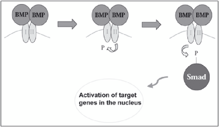
Figure 5. Mechanism of action for bone morphogenetic proteins. Upon secretion, two BMP molecules form
a dimer and bind to a complex of two transmembrane receptors, Type I and Type I. Type II receptor
phosphorylates Type I receptor, which then phosphorylates the Smad family of proteins. There proteins then
activate target genes in the nucleus including RunX2, a bone-specific transcription factor. BMPs function
as secreted signaling molecules that upon binding to a complex of transmembrane receptors, triggers a
phosphorylation cascade to activate bone specific genes in the nucleus to promote osteogenesis.
during osteogensis include osteocalcin, osteonectin, bone sialoprotein and the transcription
factor Runx2. Evidence that BMPs contribute to successful spinal fusions has been shown in
rabbits, dogs, mice and nonhuman primates.
141,180-182
However, BMPs have a short biological
half-life. Thus, coupling this treatment with the use of a biodegradable scaffold is often used.
Scaffolds used include poly-lactic acid, hydroxyapatite, de-mineralized bone matrix, and hy-
droxyapatite calcium triphosphate.
183,184
All of these examples possess the limitations outlined
in Table 1. Secondly, large quantities of BMP must be delivered in an active form to the site for
effective participation in bone healing. Finally, BMPs lack tissue specificity and thus it is diffi-
cult to control the duration and localization of their activity using the traditional carrier matri-
ces mentioned above. Gene therapy to deliver BMP to the site of desired spinal fusion can
overcome many of the obstacles faced by the traditional methods described. Introducing BMP
into cells at the fusion site will make it possible to achieve long-term controlled expression that
is tissue specific.
In experimental spinal fusions in animals, an adenoviral vector carrying rhBMP-2 is often
used. It was shown that mesenchymal stem cells transduced with the rhBMP-2 gene produced
rhBMP-2 protein in vitro and could transform into an osteoprogenitor line that can produce
bone in vivo.
185
This refers to an ex vivo method for growth factor delivery since rhBMP-2
delivery to the stem cell was performed in culture. Further, such cells were injected percutane-
ously and paraspinally at the lumbosacral junction that resulted in endochondral bone forma-
tion.
186
Proteins other than BMPs that are known to be osteoinductive such as LIM-1 (LIM
mineralization protein-1) can also be delivered to spinal fusion sites to promote osteogenesis.
159
A successful spine fusion was demonstrated; showing fusion in 100% of the sites that re-
ceived bone marrow cells that were transfected with active cDNA encoding LIM-1.
187
LIM-1
is a novel secreted protein, known to act on surrounding cells to induce bone formation. In a
rat model, posterior lateral spinal fusion was achieved following implantation of bone marrow
cells transfected with LIM-1 cDNA.
187
(Fig. 6) Therefore, genes encoding secreted osteogenic
factors are good choices for gene therapy for bone due to their ability to act on surrounding
Search WWH ::

Custom Search