Biomedical Engineering Reference
In-Depth Information
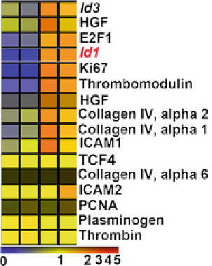
Fig. 9 Heatmap comparing
transcriptional profiles. Tree
view of the representative
Affymetrix gene expression
in the regenerative liver 48 h
after partial hepatectomy (PH
48 h) compared to that of
sham-operated mice (Sham
48 h). Note that the
upregulation of angiogenic
transcription factor Id1 is
comparable to Ki67 and
higher than E2F1, both of
which are closely associated
with hepatocyte proliferation
driven by the Id1 promoter [
71
], we found Id1 upregulation in LSECs 48 h after
PH (Fig.
8
c) which was significantly blunted in VEGFR2
fl/fl
mice (Fig.
8
d).
The liver mass recovery in Id1-deficient (Id1
-/-
) mice after PH was impaired
for 28 days and remained unchanged upon VEGF-A
164
administration (Fig.
10
a).
Furthermore, after partial hepatectomy, Id1
-/-
mice exhibited significant decrease
in mitotic BrdU
+
HNF4A
+
hepatocyte number, disrupted formation of functional
VE-cadherin
+
isolectin
+
vessels, diminished proliferation of VEGFR3
+
CD34
-
LSECs, and abnormal liver function, as evidenced by an increase in plasma bili-
rubin levels. Thus activation of the VEGF-A/VEGFR2 pathway through upregu-
lation of Id1 drives liver regeneration [
65
].
The role of Id1 upregulation in mediating the angiocrine function of LSECs on
hepatocyte proliferation was also examined by a liver SEC-hepatocyte co-culture
system. Co-incubation of isolated hepatocytes with primary LSECs led to a nine-
fold increase in hepatocyte number, which was selectively inhibited by knockdown
of Id1 in LSECs (Fig.
10
b). Conditioned medium from LSECs failed to support
hepatocyte growth, underlining the importance of cell-cell contact in liver SEC-
derived angiocrine function. Therefore, lack of Id1 results in defective inductive
function of LSECs, impairing hepatocyte regeneration.
To determine whether in vivo angiocrine effects of Id1
+/+
LSECs could initiate
hepatocyte regeneration in Id1
-/-
mice, we used the intra-splenic transplantation
approach on day 2 after PH to engraft LSECs into the Id1
-/-
liver vasculature [
72
].
GFP-marked Id1
+/+
LSECs selectively incorporated into the VEGFR3
+
sinu-
soidal vascular lumen and restored the regeneration of liver mass and SEC
expansion (Fig.
10
c). In contrast, the transplanted Id1
+/+
LSECs failed to restore
the regeneration of the Id
+/+
liver. Partial vascular chimaerism afforded by the
incorporation
of
Id1-competent
LSECs
generates
sufficient
endothelial
cell-
derived inductive signals to initiate hepatic proliferation in the Id1
+/+
liver.
Search WWH ::

Custom Search