Biomedical Engineering Reference
In-Depth Information
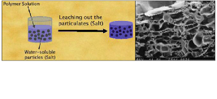
Fig. 2 Schematic of particle leaching fabrication method. The polymer solution is placed in a
mold with some sort of porogen (i.e. salt) and then a scaffold is formed. The porogen is then
washed out of the scaffold, leaving a network of interconnected pores [
31
](left). SEM images of
porous ceramic scaffold fabricated through particle leaching method through dissolution of the
porogen [
32
](right)
Earlier studies discovered that pore diameter and biodegradability of the scaffold
significantly influence the cellular efficacy to construct vascular networks within the
scaffold and also bridge new blood vessels with pre-existing ones [
10
]. For example,
human embryonic endothelial cells or human umbilical cord vein cells (HUVEC's)
were incorporated into a myoblast-laden biodegradable PLGA scaffold, in order to
crate vascularized muscle [
11
]. Micropores were incorporated into the scaffold using
the salt leaching method [
12
]. The resulting scaffold pore diameters range from 225
to 500 lm with a porosity of 93 %. Endothelial cells secreted VEGF and PDGF
within the pores and organized into tubular structures in vitro. Furthermore, in vivo
studies, where the scaffold was implanted subcutaneously, showed that the prevas-
culature within the scaffolds integrated with host vasculature in SCID mice [
11
].
Alternatively, porous PLGA scaffolds were prepared using the gas foaming/
particle leaching method. The copolymer, mixed with 100 mg of NaCl, was
compressed into a disk form, and the disk was placed in a pressure chamber filled
with carbon dioxide. After the gas was partially dissolved into the polymer disk,
the chamber was depressurized to convert the carbon dioxide smeared into the
matrix into a gaseous form and subsequently generate the pores in the polymer
disks. Further leaching of NaCl in aqueous solution created the microporous
scaffold porosity of 95 % with an average pore size of 190 lm. The human micro
vascular endothelial cells (HMVECs) were incorporated into scaffolds by dropping
cells in suspension into the pores. These scaffolds were capable of developing
mature blood vessels via vasculogeneis within the scaffold (Fig.
3
), as demon-
strated with the scaffold subcutaneously implanted into SCID mice [
13
].
In addition, mesenchymal stem cells transfected with genes encoding VEGF
and bFGF were used by loading them in a porous biodegradable collagen and
glycosaminoglycan scaffold with the commercial name of the Integra
matrix. The
matrix is composed of cross linked bovine collagen and glycosaminoglycans [
14
].
The resulting matrix has a porosity of 98 % and pore sizes ranging from 30 to
120 lm. Cells seeded in the matrix actively secreted exogenous VEGF and bFGF.
The cell seeded scaffold was tested in vivo using a skin defect model where a
Search WWH ::

Custom Search