Biomedical Engineering Reference
In-Depth Information
A
B
(a)
(a)
(b)
C
(a)
(b)
(b)
(c)
(d)
(c)
(c)
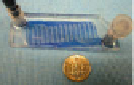
Fig. 2 Preformed microvascular networks: A (a) Macroscopic view of the microfabricated
PDMS network. (b) Endothelial cells immediately after seeding onto the pre-fabricated networks.
(c) Endothelial cells lining the networks after monolayer formation [
77
]—Reprinted with
permission from Springer. B Formation of tubes that incorporate endothelial cells and fibroblasts.
(a) Schematic diagram showing the fabrication of microfluidic gels. Sequential introduction of
Pluronic and liquid gelatin into the channels, and gelation, yielding a gelatin mesh that could be
separated from the channels. (b) Collagen gel with embedded fibroblasts. (c) Collagen gel with a
monolayer of endothelial cells lining internal channels. Inset, Hoechst-stained microvascular
network [
78
]—Reproduced by permission of The Royal Society of Chemistry. C (a) Schematic of
gel substrate with embedded perivascular cells (PCs) and a channel for endothelial cell seeding.
(b) Phase-contrast image and (c) corresponding fluorescence image of the center plane of the tube
with PCs embedded within the collagen matrix. (d) Fluorescence image after 20 min of perfusion
with fluorescently labeled BSA. Scale bars = 100 lm[
80
]—Reprinted with permission from
Elsevier Ltd
by the need for vascular networks in organs being developed for engineered tis-
sues. One of the first demonstrations of microvessel growth in PDMS channels was
published in 2002 [
76
,
77
] (Fig.
2
A), demonstrating that the vessels could be
maintained for up to 2 weeks under flow conditions in channels as small as 35 l.
Of course, the use of these ''engineered'' vascular networks would require that
either the channels be cast in a hydrogel that could be seeded with cells, as has
recently demonstrated [
78
,
79
] (Fig.
2
B), or that the networks be removable from
the mold without causing irreparable damage.
An alternative ''engineered'' approach was developed by Tien and colleagues
[
80
,
81
] (Fig.
2
C). In their approach, a cylindrical channel was cast around a rigid
needle in a hydrogel such as type I collagen. Once gelled, the needle was removed,
leaving an open cylinder into which endothelial cells could be seeded, forming
confluent EC-lined lumens after about 2 days in culture. These vessels could sub-
sequently be perfused, and in doing so, became stabilized through a combination of
physiological levels of fluid shear stress and hydrostatic pressure [
81
]. Functional
assays were performed, demonstrating levels of permeability for albumin compa-
rable to those observed in vivo (*2 9 10
-7
cm/s) and well-defined, continuous


Search WWH ::

Custom Search