Biology Reference
In-Depth Information
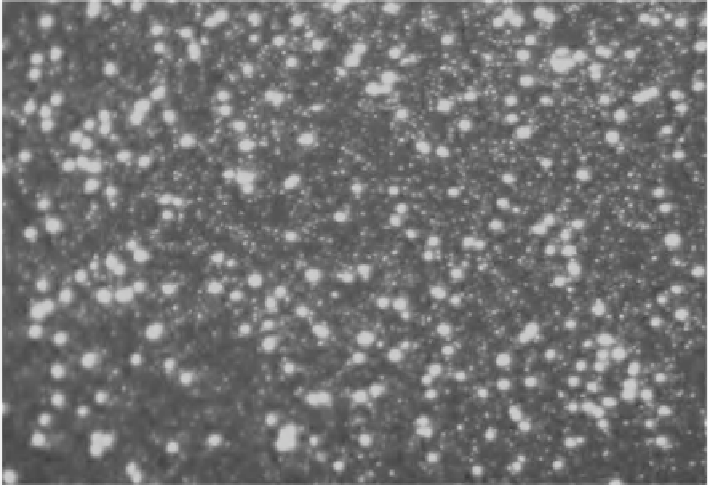
Figure 8:
An image from an epifl uorescence microscope of seawater stained with the dye SYBR Green to reveal the bacterial
cells and smaller viral particles. With the kind permission of N. H. Mann Department of Biological Sciences, University of
Warwick, Coventry, UK.
[Mann, N. H. (2005)
PLoS Biol
3(5):
e182. doi:10.1371/journal.pbio.0030182]. doi: 10.1371/journal.
pbio.0030182.g001. Image courtesy Jed Fuhrman, Department of Biological Sciences, University of Southern California,
MC0371, Los Angeles, CA 90089, USA.
Color image of this figure appears in the color plate section at the end of the topic.
of 25 mm Anodisc
TM
membranes with a built-in support disc has been practised. But Budinoff
et
al
. (2011) advocated the use of 13 mm Anodisc
TM
or Nucleopore
TM
membranes (with pore sizes
0.015-0.030 µm) without a support disc. Hennes
et al
. (1995) used fl uorescently stained viruses as
probes to detect specifi c bacteria and cyanobacteria. Bettarel
et al
. (2000) compared fi ve different
methods for the enumeration of marine viruses. These are TEM, and epifl uorescence microscopy
with different fl uorochromes such as DAPI, YO-PRO-1, SYBR Green I. Of these the use of YO-PRO-1
in combination with epifl uorescence microscopy proved to be the best for the enumeration of T7
virus particles from cultures as well as viruses from different freshwater ecosystems that differed
in their trophic levels. Marie
et al
. (1999) enumerated a marine virus PpV-01 in culture infecting
Phaeocystis pouchetii
as well as natural samples through fl ow cytometry, a technique that has been
used for counting the number of bacterial or cyanobacterial (
Prochlorococcus
or
Synechococcus
) cells
from marine environments (Fig. 9). After fi xing the samples with 0.5 % glutaraldehyde, the samples
were deep-frozen and in presence of SYBR Green I, the enumeration of viruses has been done by fl ow
cytometry (Fig. 10). However, the titres of phages were found to be slightly on higher scale when
compared to epifl uorescence microscopy or by TEM. They also found that the viruses displayed a
depth-dependent profi le from the Mediterranean Sea as that of the cells of the host. A disadvantage
with SBYR Green I is that its fl uorescence fades within 30 seconds under some conditions. As a result
of which one is bound to use high concentrations of SBYR Green I and also employ an anti-fading
mixture (Noble and Fuhrman, 1998). That is why some workers preferred to use SBYR Gold as
the fl uorochrome instead of SBYR Green I. In this connection, the work of Chen
et al
. (2001) merits
mention because they employed digital image analysis and fl ow cytometry to enumerate marine