Biology Reference
In-Depth Information
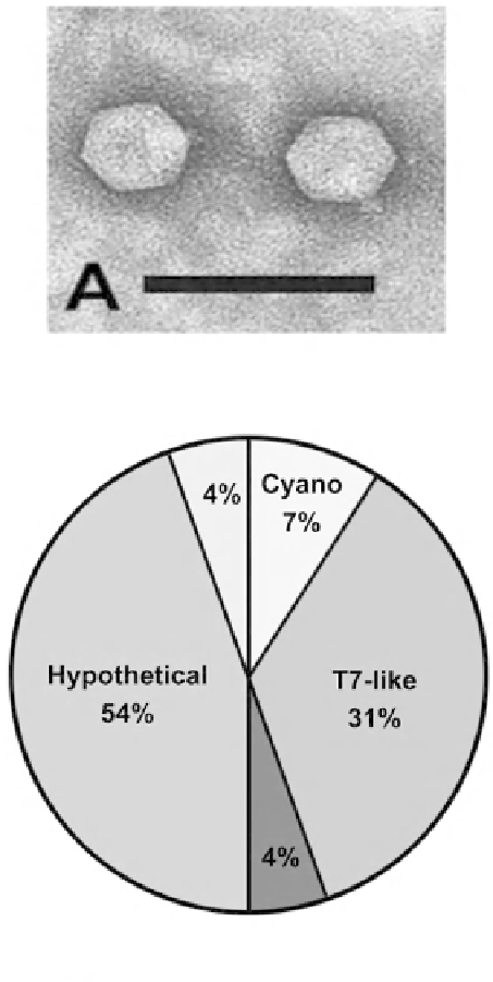
B
Figure 7:
Features of the
Prochlorococcus
Podovirus P-SSP7 (A) Electron micrograph of negative-stained podovirus P-SSP7.
Note the distinct T7-like capsid and tail structure. Scale bar indicates 100 nm. (B) Taxonomy of best BLASTp hits for P-SSP7.
Each predicted coding sequence from the phage genomes was used as a query against the nonredundant database to identify
the taxon of the best hit. Blue slices indicate phage hits, while yellow slices indicate cellular hits. With the kind permission of
S. W. Chisholm, Department of Biology, Massachusetts Institute of Technology, Cambridge, Massachusetts, USA. [Sullivan
et al.
(2005)
PLoS Biol
3
(5):
e144. doi:10.1371/journal.pbio.0030144] doi:10.1371/journal.pbio.0030144.g001.
Color image of this figure appears in the color plate section at the end of the topic.