Biology Reference
In-Depth Information
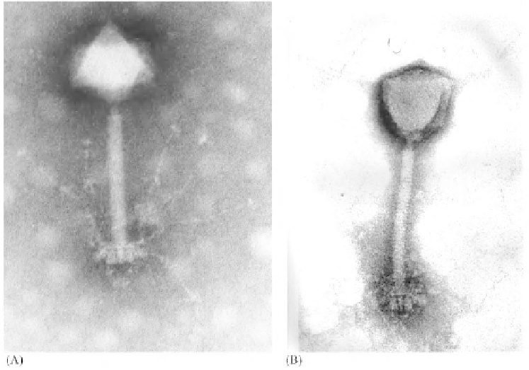
Figure 4:
(A) Transmission electron microscope image of the
Synechococcus
Phage S-PM2. (B) Negative (phosphotungstate-)
stained particle of S-PM2. x 297,000. Picture (A)
with the kind permission of N. H. Mann, Department of Biological Sciences,
University of Warwick, Coventry, UK.
[Mann, N. H. (2005)
PLoS Biol
3(5):
e182. doi:10.1371/journal.pbio.0030182] Doi:10.1371/
journal.pbio.0030182.g002.
Images (A) and (B) courtesy H.-W. Ackermann, Department of Microbiology, Immunology and Infectiology, Faculty of
Medicine, Laval University, Vandry Pavilion, Avenue de la Médecine, Quebec, QC, G1K 7P4, Canada.
and cyanosiphoviruses (from Bermuda and English Channel) resembled each other. There is a
limited degree of homology among all cyanophage DNAs as revealed by Southern hybridization.
The structural protein in each case was ~53 to 54 kDa.
Lu
et al
. (2001) isolated and characterized a number of cyanophages from three Georgia's coastal
river (Savannah, Altamaha and Satilla) estuaries by selecting four phycoerythrin (PE)-containing
strains (WH7803, WH7805, WH8108, and WH81103) and seven phycocyanin (PC)-containing
(WH8101, WH8007, WH5701, CCMP1628, CCMP1629, CCMP1630 and CCMP1632) strains of
Synechococcus
sp. as the hosts for isolation. Cyanophages P3 (isolated on WH7805), P5 and P16
(isolated on WH 7803) cross-infected majority of PE- and PC-containing strains and so had broad
host range. Cyanophages P68 and P76 (isolated on PC-containing strain WH 8007) exhibited a very
narrow host range as the former cross infected only WH 8101 while the latter did not cross infect
any other strain. Cyanophages (P2, P39, P53, P56, P71, P73, P79, P81, P82 and P83) isolated on WH
7805 cross infected other PE strains such as WH7803 and WH8108. Of the cyanophages P8, P12 and
P14 isolated on PC-containing strain WH8101, only P8 and P14 cross-infected WH7803 and WH 7805
whereas P12 did not cross infect any other strain. Cyanophages P71, P72, P73, P79, P81, P82 and P83
isolated on PE-containing strain WH7805, all cross infected WH7803 and WH8108. Morphologically
80% of the cyanophages belonged to the family Myoviridae (Table 8).
Northern waters of Moreton Bay, Queensland, Australia supported expansive bloom of
Lyngbya
majuscula
that produces toxic substances responsible for dermatitis and asthma-like symptoms.