Biology Reference
In-Depth Information
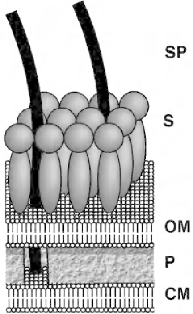
Figure 2:
Diagram imaging the present knowledge of the cell envelope structure of
Synechococcus
. Present observations
taken together suggest that the spicules (SP) extend through the surface layer (S) and outer membrane (OM) to contact the
cell membrane (CM) (as shown in the cutaway of the peptidoglycan layer (P)).With the kind permission of A.D.T. Samuel,
Rowland Institute for Science, Cambridge, Massachusetts, USA and Department of Molecular & Cell Biology, Harvard
University, Cambridge, Massachusetts, USA [Samuel
et al
. (2001)
BMC Microbiol
.
1:
4. doi: 10.1186/1471-2180-1-4].
gradient did not show such a correlation suggesting that the sodium motive force provides the direct
source of energy for swimming in this organism. On the basis of the requirement of calcium for
swimming, Pitta
et al
. (1997) concluded that the cell surface experiences longitudinal or transverse
waves and the wave motion is suggested to be coordinated or driven by the changes in calcium
concentration. Calcium depolarization is suggested to cause a local swelling of the cell surface which
is responsible for the production of waves (Ehlers
et al
., 1996). Whether it is the sodium motive force
or calcium that provides the energy for thrust, according to Samuel
et al
. (2001) the spicules noted
by them in the cell envelope of
Synechococcus
sp. strain WH8113 are well positioned to transduce
energy at the cell membrane into mechanical work at the cell surface.
C) Twitching movements
The presence of pili or fi mbriae on the cell surface is a characteristic feature of a number of eubacteria.
The pili present on
E
.
coli
have been classifi ed into four categories, i.e. Type I, II, III and IV on the basis
of their diameter, number and distribution (Hayes, 1968). In cyanobacteria, the presence of pili-like
structures on the cell surfaces of certain unicellular and fi lamentous forms has been demonstrated