Biology Reference
In-Depth Information
(A) (B)
(C) (D)
(E) (F)
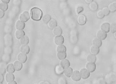
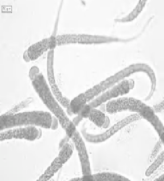
Figure 1:
Light micrographs of
Anabaena
sp. (A),
Nostoc
sp. (B),
Calothrix
sp. (C),
Anabaenopsis circularis
(D),
Scytonema
sp. (E) and
Anabaena
sp. (F) showing intercalary heterocysts with two polar nodules (in A,B, D, E and F) and
a single basal heterocyst with
a single polar nodule (in B). The presence of a pair of heterocysts is characteristically seen in (D). The polar nodules can be seen
clearly in (F). Magnifi cation bar in the pictures represents 5 µm (A) and 10 µm (C, D and E). Pictures A,C and E courtesy G.L. Tiwari,
Department of Botany, University of Allahabad, Allahabad-211002, India.
Picture B courtesy Isao Inouye (Uinversity of Tsukuba),
Mark Schneegurt (Wichita State University) and Cyanosite (www-cyanosite.bio.purdue.edu). Picture F courtesy A. L. Baker,
Department of Biological Sciences, 124 Spaulding Life Sciences, Academic Way, University of New Hampshire, Durham,
NH 03824 USA. http://cfb.unh.edu/phycokey/phycokey.htm.