Biology Reference
In-Depth Information
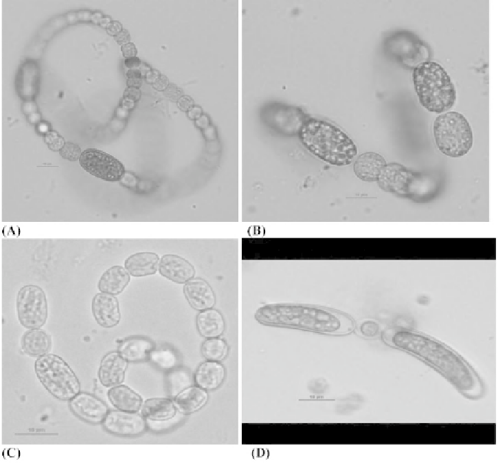
Figure 1:
Development of akinetes in species of
Anabaena
.
A
.
circinalis
(A),
A
.
crassa
(B),
A
.
lamermannii
(C) and mature
akinetes of
A
.
lamermannii
(D). In (A) and (B) development of akinetes a little away from heterocysts can be noted whereas
in (C) and (D) the development of akinetes on either side of the heterocyst can be noticed. The magnifi cation bar in the
fi gures represents 10 µm. Pictures courtesy Mark T. Aubel, GreenWater Laboratories/CyanoLab, 205, Zeagler Drive, Suite
302, Palatka, Florida 32177, USA. (http://www.greenwaterlab.com/photo_algal.htm).
number of akinetes formed in the majority of genera is quite large as for example in certain species of
Anabaena,
akinetes are formed in strings or chains as all vegetative cells get transformed into akinetes
(Fig. 2 C). In
A
.
lammermanni
the akinetes are formed in large numbers and they settle to the bottom
of the water body (Fig. 4 A). However, few akinetes are formed in
Cylindrospermum
(subterminally
beside a heterocyst on either side of fi lament) and
Gloeotrichia
(where only one subterminal akinete
is present in each fi lament). In certain species of
Anabaena
(
A
.
circinalis
),
Anabaenopsis
(
A
.
raciborskii
)
two akinetes differentiate in the middle of the trichome. The akinetes formed in case of
Anabaena
cf.
macrospora
appear to be the largest in size (Fig. 4 B).
3) Structure
:
In the light microscope, the akinetes appear as thick-walled and granulated structures
very much larger than their antecedent vegetative cells. They appear as dark brown or blackish
structures. The ultrastructure of akinetes revealed that photosynthetic lamellae remain intact and the
presence of glycogen, cyanophycin, lipid granules and polyhedral bodies has been demonstrated.