Biomedical Engineering Reference
In-Depth Information
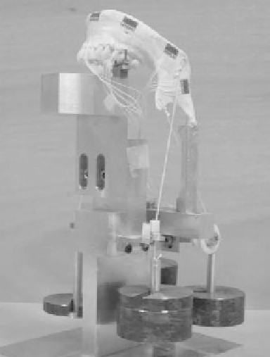
Fig. 12
Synthetic mandible
with locations of rosette
strain gauges. Bone strains
were measured with four
gauges glued onto the
external mandible surface
The FEM used in the present study has been validated [
58
]. The model was
experimentally validated with tri-axial strain gauges (CEA-06-062UR-350, Vishay
Electronic GmbH, Germany) and the fiber brag sensors technique. To validate the
accuracy of the FE model, simplified boundary conditions were used, as shown
in the apparatus Fig.
12
. In the model, validation strain was only applied to two
muscles in each side, with a lower magnitude 5 and 10 N. Four nylon wires were
glued at the anatomical insertions of the masseter and temporal muscles and used
to load the mandible by placing weights at the end of them. In this validation
the material properties of the mandible in FEM was the Sawbones with 460 MPa
Young's modulus.
In this experiment, three different mandible support situations were considered,
incisor, canine and molar teeth. Four load situations were analyzed, Load 1 applied
masseter muscle with 5 N, Load 2 the same muscle with 10 N, Load 3 configuration
includes the master muscles with a force of 10 N and the temporal muscle force of
5N.Load 4 configuration included forces of 12 N, 6 N, 2 N respectively for the right
masseter, right temporal and left masseter muscles. No force was considered for the
left temporal (Tleft) muscle. The experimental and numerical results are presented
in Fig.
13
for canine teeth support.
The results revealed a good correlation between FEM and experimental results
and considering all FE and measured strains, the correlation value R
2
and slope
of the regression line are 0.98 and 0.93 for the maximum and minimum principal
strains presented in Fig.
14
.