Biomedical Engineering Reference
In-Depth Information
Fig. 8
Determining the muscle sections
be known. Several parallel and close recordings, orthogonal with this longitudinal
muscle axis, were then made.
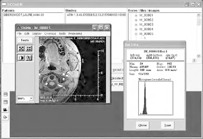
The digitized images were analyzed using image processing software (Osiris,
Hopitaux Universitaires de Geneve, Switzerland). A module makes it possible to
select one area of the image and characterize it. Thus this area of interest can, for
example, delimit the two masseters (Fig.
8
). The software revealed here a total area
of the section of nearly
454 mm
2
.
2.2.4
Evolutions of the Protocol and Accuracy of the First Results
The contribution of the posterior temporal muscle remained weak compared to that
of the other two muscle bundles (medial, and anterior): dividing the temporal muscle
into two bundles therefore seemed sufficient. The objective was first to improve the
precision of the results and to circumvent the difficulty of vectorizing this fan-like
muscle. Because of the volunteers' hair, surface electrodes could be used only for the
anterior bundle of the temporal muscle. This suppression of four thread electrodes
was interesting, however, because it simplified the experimental procedure and
reduced the pain experienced by the volunteers. Reducing volunteer pain will also
probably improve the accuracy of results.
Tab le
1
presents the magnitudes of the forces for the six pairs of jaw-closing
muscles. In each position, three tests were carried out for each of the four volunteers.
Averages were calculated on the basis of 12 values.