Biomedical Engineering Reference
In-Depth Information
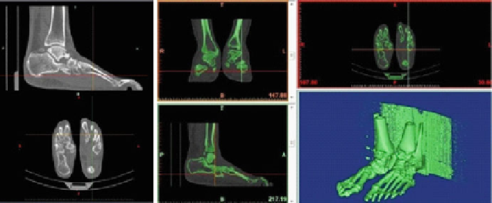
CT from an individual feet and software Mimics
®
Fig. 8
3.2
3D Foot Models
In the attempt to find and develop a reliable and efficient method to build 3D
anatomical structures for reverse engineering, a methodology including the use
of different interconnected software, was validated. It consists on using medical
imaging files to develop the 3D model, using design software, for Finite Element
Method (FEM) simulation, making possible to obtain a 3D foot model of each
individual. There are similar methodology studies, starting from CT scans and using
CAD software (CATIA
®
) to develop a model ready to be tested on FEM simulation
(Abaqus
®
) in order to find either, strain and stress on the foot, or to see stress
differences considering specific tendons and muscles or even stress for different
foot positioning for 2D and 3D foot models [
1
,
9
,
10
,
18
,
31
].
These studies showed it is possible to take a file from medical images, CT, MRI
or even Micro-CT scanner, even though the latter aren't point clouds but rather a set
of 2D slices which are unified to produce a 3D representation. During a subject
CT or MRI exam, it is important that each foot is in the position adequate for
the study objective, allowing for original tissue assembly and avoiding wrong 3D
objects manipulation. For example, to study the foot evolution during gait, which
is obviously done in the standing-up position, when doing a CT scan, and because
this medical exam is done lying-down in a table, it is important to have the patient
feet vertically aligned as they would be in the focused movement. As a curiosity, for
high heels studies subject's feet must have the calcaneous slightly raised.
Using software like Mimics
®
, and taking patient feet medical images (DICOM
files) it is possible to get a single file with the corresponding point clouds, according
to tissue density (Fig.
8
). Therefore, one can get 3D objects corresponding to bones,
with or without cartilage, and soft tissues, with or without differentiating muscles
tissue, fat tissue, skin tissue, using density segmentation techniques related to the
different masks provided by the software (Fig.
9
).
For our study mesh clouds were divided into two groups: bone structure (that
includes bones and cartilage) and soft tissue. They were then exported as STL files,