Biomedical Engineering Reference
In-Depth Information
0
ILM
−
500
1000
−
RPE
−
1500
−
2000
−
3000
−
2000
−
1000
0
1000
2000
3000
Lateral Direction [
µ
m]
200
100
0
0
1800
−
2000
−
200
−
400
−
600
−
800
−
1000
−
1200
−
1400
−
1600
−
Axial Direction [
µ
m]
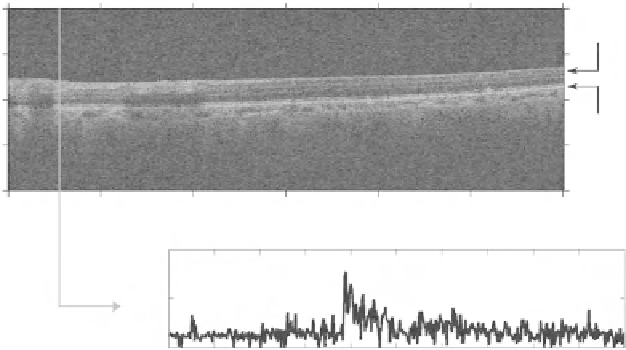
Fig. 1
Optical coherence tomography example of a B-scan (
top
) and an A-scan profile (
bottom
)
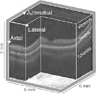
Fig. 2
Volumetric OCT data
shown over an eye fundus
reference
In this work the high-definition spectral domain Cirrus OCT (Carl Zeiss Meditec,
Dublin, CA, USA) was used.
This retinal imaging system allows for an acquisition scan of 200
200
1;024
or 512
1;024 voxels for the lateral, azimuthal and axial directions, respec-
tively, with a depth and lateral resolutions of 5 and 20 m in tissue, respectively.
This volumetric data is obtained from a 6;000
128
6;000
2;000 m
3
volume of the
human macula (Fig.
2
).
OCT readings result from reflections and/or light scattering due to refractive
index changes along the light path and are therefore dependent on the content and
structure organization of the eye.
OCT has been used to assess structural information from the eye fundus, in vivo,
allowing to identify and compare retinal changes in different instants of time.