Biomedical Engineering Reference
In-Depth Information
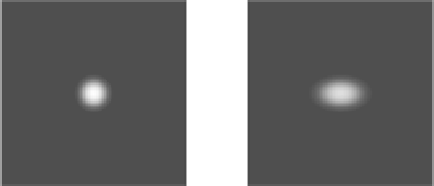
Fig. 12
Illustration of the
image blurring caused by
movement of a point source.
(
a
) Acquisition of a static
point source. (
b
) Hypothetical
situation of a point source
with a given periodic
translation movement
a
b
depended on the time and nature of the motion and also on the number of camera
heads used, e.g.: for deviations of less than 1 pixel no perfusion defects were
detected (assessed by QPS
4
), but for the deviations of 3 pixels the defects detected
corresponded to 8.1% for a camera of one head and 11.8% for a camera with
two heads.
Regarding PET, Osman et al. investigated the effect of motion in PET/CT studies
finding deviations between PET and CT in the localization of lesions. According
to this study, the localization deviations between the two techniques, which are
afterwards fused, can result in some cases (2%) in gross lesions localization errors.
Moreover, the need for attenuation correction in PET from the CT images cause
several artifacts when there is patient motion [
34
-
36
]. It was also shown that
respiratory motion causes a reduction in the accuracy on the determination of
volume and activity of lung lesions when examined with
18
F-FDG PET [
37
,
38
].
Studies performed in other areas [
39
-
42
], particularly in radiotherapy, report
an increase of the volume and errors in the localization of tumors that lead to
inadequate planning.
3.2
Methods for Detecting Motion
Motion detection is the precursor of the correction and is a key step for the success
of any technique to compensate for motion effects. There are numerous forms to
detect motion that could be classified into two groups: techniques that use directly
the scintigraphic images (data driven) and techniques that utilize external sensors to
detect the motion.
4
Quantitative Perfusion SPECT.