Biomedical Engineering Reference
In-Depth Information
3.4
Examples
Reconstruction of the different cardiovascular structures can be performed using
the generic flow process in Fig.
3.11
. Segmentation algorithms form the core step in
extracting the region of interest. However instead of presenting the same procedures
in this section, alternative methods are presented.
3.4.1
Abdominal Bifurcation
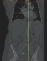
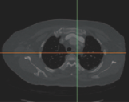
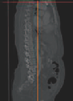
The DICOM images of a CT scan from a 61-year-old female patient is shown in
Fig.
3.12
in three planes of view: coronal, axial, and sagittal. The original CT scan
images comprised of 955 contiguous slices with a slice increment of 0.625 mm.
The scan image had a resolution of 512 × 512 pixels, with a pixel size representing
0.703 mm.
Thresholding segmentation is first applied by defining the minimum and maxi-
mum pixel greyscale values based on the Hounsfield scale (set to 168 and 596
respectively—see Sect. 3.2) to extract the artery passageway. The thresholding seg-
mentation extracts the pixels within the defined limits and is coloured in green in
Fig.
3.13
. However this will also include additional side artery branches and other
tissue that exhibit pixels within the threshold range. In such a circumstance, manual
operation is needed to remove these additional pixels that are not part of the region
of interest.
The final segmented volume is then extracted from the threshold segmentation.
A preliminary three-dimensional surface geometry is reconstructed based on the
exported CAD file as an .
stl
format. During the CAD conversion, a large amount of
noise is present and the preliminary 3D geometry requires further refinements. The
preliminary model and the refined model are shown in Fig.
3.14
.
&RURQDO
D[LDO
VDJJLWDO
Fig. 3.12
Orientation assignment of the imported scanned images
Search WWH ::

Custom Search