Biomedical Engineering Reference
In-Depth Information
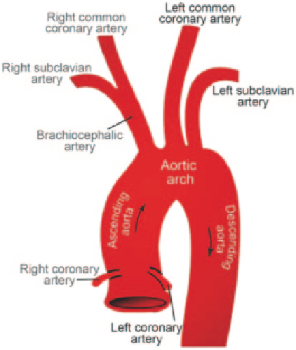
Fig. 2.10
Components of the
aortic arch. The
right and left
common coronary arteries
(
CCA
) branch from the aortic
arch. The
right CCA
extends
from the brachiocephalic
artery
known as the trunk or innominate artery, extends from the first branch of the aortic
arch, and divides into the right common carotid artery and the right subclavian
artery. It connects to the mediastinum that supplies blood to the head and neck as
well as the right arm.
The ICA and ECA branches supply blood to different organs through arteries at
their downstream. In particular, the brain and eyes located at the downstream of ICA
branch are the most active and important part of the human body, which consume
a high quantity of oxygen and require a high volume of blood supply per unit time.
Therefore the blood flow volume through the ICA branch is greater than that of the
ECA.
Common Carotid Artery (CCA)
The Common Carotid Artery ascends through the
superior mediastinum
2
anterolaterally in the neck and lies medial to the jugular
vein
3
. The two CCA are not symmetrical, with the left artery having greater length
than the right artery. This accounts for the longer path from the aortic arch. The
carotid artery, jugular vein, and vagus nerve are enclosed in the carotid sheath. The
CCA bifurcates into the internal carotid artery and the external carotid artery at the
superior border of the thyroid cartilage with inter-individual variations in terms of
angle of bifurcation and asymmetry. The diameter of the CCA in adults ranges from
0.2-0.8 cm with an average value of 0.7 cm (Xu 2002).
2
The
mediastinum
is the central compartment in the thorax that contains a group of structures that
includes the heart, the esophagus, the trachea, and the lymph nodes of the central chest.
3
The
jugular vein
is part of the venous network of vessels that bring deoxygenated blood from the
head back to the heart via the superior vena cava (see Fig.
2.2
).
Search WWH ::

Custom Search