Biomedical Engineering Reference
In-Depth Information
coronary arteries are analogous to the organs of the human body and the cardiovas-
cular network of arteries.
2.2
Physiology of the Cardiovascular System
2.2.1
Anatomy of the Heart
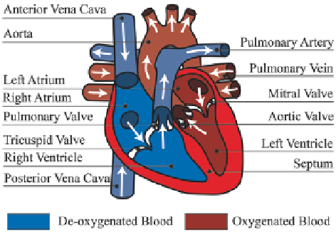
The heart supplies oxygenated blood to the rest of the body and then transports
the de-oxygenated blood to the respiratory system for oxygen replenishment
(Fig.
2.4
). The heart comprises the right ventricle and left atrium, separated by
a partition septum. Each half consists of two chambers; a thin-walled atrium
and a thick-walled ventricle. The atria receive blood from the veins, while the
ventricles pump blood out of the heart and through the circulatory system. The
right atrium is the upper right chamber of the heart collecting de-oxygenated
blood from the vena cava, and then passes it via the tricuspid valves. This goes
into the right ventricle for pumping into the lungs through the pulmonary valve
and via the pulmonary artery for oxygenation. The oxygenated blood returns via
the pulmonary vein into the left atrium, which then pumps the blood into the left
ventricle through the mitral valve. The left ventricle is the strongest chamber
of the heart that supplies the oxygenated blood to the rest of the human body
via the aorta after passing through the aortic valves. The valves in the tricuspid,
Fig. 2.4
Cardiovascular circulation of the heart. The heart comprises the
left and right atria
,
which are responsible for collecting de-oxygenated and oxygenated blood from the vena cava and
pulmonary vein respectively. The
right ventricle
is a heart chamber that pumps de-oxygenated
blood to the respiratory system, and
left ventricle
is the most muscular chamber that pumps the
oxygenated blood to all parts of the body. Heart valves are present at the connections of the atria
and ventricles, as well as the pulmonary artery and aorta to achieve a single flow direction circuit.
The
white arrows
indicate the direction of blood flow
Search WWH ::

Custom Search