Biomedical Engineering Reference
In-Depth Information
aortic dissection, and also promotes the closure of the dissection layer and improve
blood perfusion of the lower limbs. This treatment is an effective procedure to treat
DeBakey III aortic dissection.
8.8
Mitral Valve Dynamics
8.8.1
Introduction
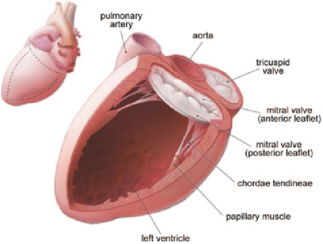
The mitral valve is composed of the anterior and posterior leaflets, the chordae ten-
dineae, and the papillary muscles; separating the left ventricle and left atrium in the
heart. It behaves as a valve, controlling the flow of blood. During diastole it allows
blood flow from the left atrium to the left ventricle. During ventricular systole, the
mitral valve closes and prevents backflow of the left atrium. The mitral valve action
is caused by blood flow associated with pressure differences over the leaflets and
leaflet muscle fibre activation resulting from a complex interaction of wall tension
and flow pattern in the atrium and ventricle compartments (Skallerud et al. 2011;
Fig.
8.68
).
Mitral valve function requires the coordinated action of its interrelated compo-
nents, and alterations in its structure caused by remodeling, stenosis, or weakening
leading to regurgitation, may lead to eccentric ventricular hypertrophy, pulmonary
Fig. 8.68
Mitral valve is composed of the anterior and posterior leaflets, the chordae tendineae,
and the papillary muscles. (adapted from Wikicommons)
Search WWH ::

Custom Search