Biomedical Engineering Reference
In-Depth Information
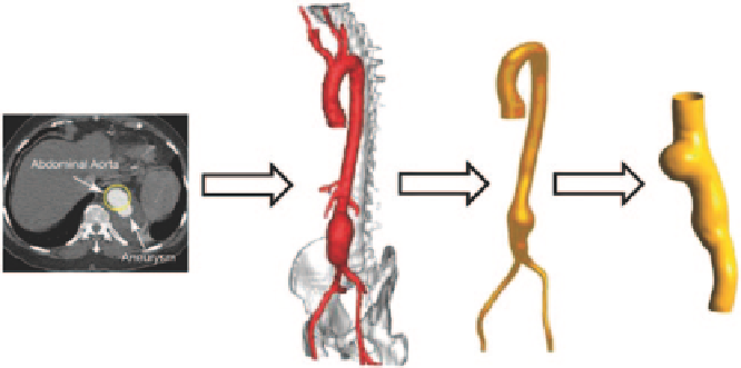
Fig. 8.51
Reconstruction of an abdominal aortic aneurysm based on multi-slice CT angiography
8.6.1
Geometric Models and Material Properties
An anatomical realistic model was reconstructed from multi-slice CT angiography
of an abdominal aorta with a moderate aneurysm. The reconstruction technique has
been described in the previous chapter. In this case study, only the artery vessel with
aneurysm (Fig.
8.51
) was selected for numerical simulation.
The aneurysm wall was assumed as hyperelastic, homogenous, incompressible
and isotropic. Similar to the carotid artery vessel, a widely accepted strain ener-
gy density function, the Mooney-Rivlin model was used to express its elasticity
(see Eqn 8.1). The model parameters were set to
C
10
= 0.070 MPa,
C
20
= 3.2 MPa,
C
21
= 0.0716 MPa,
D
1
= 0.1 MPa, and
C
ij
= 0 MPa for the others. The arterial ves-
sel wall thickness is 0.5 mm thickness. Using multi-blocking with O-grid strategy,
the computational fluid domain was meshed into structured elements, and uniform
thickness was assigned on the fluid domain surface through extruding the arterial
inner wall outwards. All mesh results are shown in Fig.
8.52
. Again, mesh refine-
ment was imposed at the near wall regions of the fluid domain to increase the spatial
resolution.
Representative flow waveforms shown in Fig.
8.53
were used as boundary con-
ditions, at the inlet a pressure inlet was assigned, while at the outlet, a velocity
outlet boundary condition was applied. Both inlet and outlet were constrained with
fixed support (Fig.
8.54
), and no movements and deformation were allowed at these
locations.
8.6.2
Haemodynamics Inside the Abdominal Aortic Aneurysm
Velocities at the cross-sectional plane of the aneurysm were plotted for the CFD and
FSI modelling approaches (Fig.
8.55
). The CFD model using a rigid wall assumption
Search WWH ::

Custom Search