Biomedical Engineering Reference
In-Depth Information
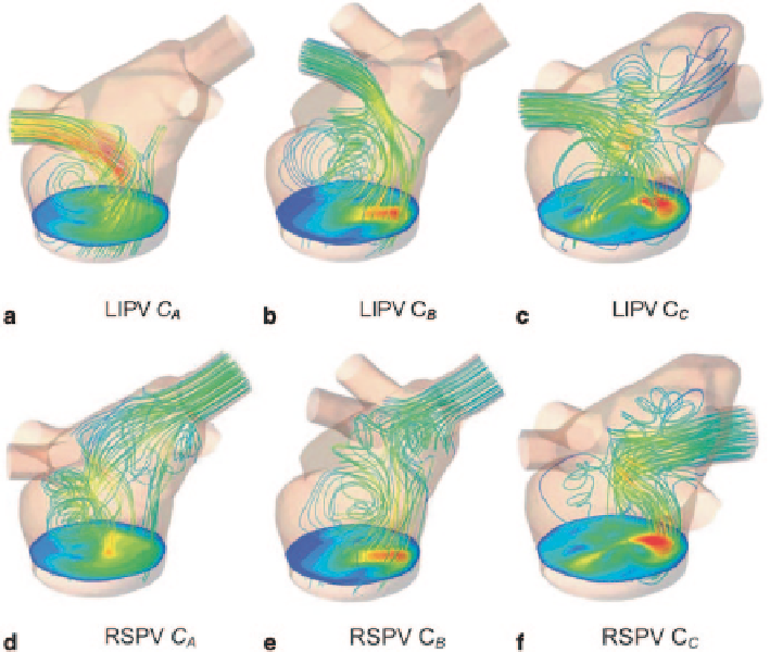
Fig. 7.41
Streamlines from the LIPV (left inferior pulmonary valve) and RSPV (right superior
pulmonary valve) are plotted for each configuration at their maximum velocity. The maximum
velocity occurs at
t
= 160
ms
,
t
= 205
ms
and t = 190
ms
for
C
A
,
C
B
and
C
C
, respectively
7.6.4
Closure
In this section we presented three 3D CFD simulations focusing on the intra-atrial
flow and the resulting mitral plane velocity profile during left ventricle diastole. The
anatomically based 3D geometries of the left atrium and the pulmonary veins were
obtained from MRI recordings of a young healthy adult. The entry locations of the pul-
monary veins were different in the three models. Four jets enter the asymmetric atrium,
and the resulting flow field is therefore complex. The results clearly illustrate that the
locations of the pulmonary valves have a significant impact on the intra-atrial flow.
When comparing the flow field in the three models, the results clearly illustrate
that the pulmonary veins have a significant impact on the intra-atrial flow and the
final mitral plane velocity profile. Because the interpatient variability in venous
number and branching patterns is large, the mitral plane velocity profile should be
considered as a subject-specific property. Therefore, we suggest that in order to
Search WWH ::

Custom Search