Biomedical Engineering Reference
In-Depth Information
7.4.6
In-Vitro Flow Measurement of an Aneurysm, Based on PIV
Fluorescent stereo PIV can be used to perform flow measurement of the aneurysm
for flow comparison with the CFD simulation (Takahashi et al. 2011). Fig.
7.28
,
shows a DS20 Laser with a 1 kHz double-pulse, 527 nm wavelength at 5 mJ/pulse
and thickness of 80 ~ 100 μm used. The fluid medium is a glycerol and water mix-
ture at 3:2 ratio (by weight). This results in a density of 1.16 kg/m
3
, viscosity of
5.3 cP at 37°, and refractive index of 1.409 ± 0.002 (532 nm at 37°). The fluores-
cent particle, FLUOSTAR
®
, which has a density of 1.1 kg/m
3
and diameter of
13 μm, was added as motion tracking particles at a concentration of 1.1 g/L. The
measurement used an interrogation size of 16 × 16 pixel
2
, with spatial resolution
of 110 μm, and laser light of 80 ~ 100 μm thickness, and scanning plane of 80,
phase-locked time-binning of 30 ms. Measurements were taken at peak time phase
resulting in Re = 450.
The residual neck size, and vessel channel geometry affects the flow field inside
the aneurysm, and consequently influences success of the aneurysm treatment. Ide-
ally the aim is to achieve reduced inflow and intra-aneurysmal velocities (Byun and
Rhee 2003). Stereo PIV allows 3D measurement and characterization of abnormal
flows such as an impinging jet in a near-wall region of the aneurysm (Fig.
7.29
). By
correlating with pathophysiology, early prevention of aneurysmal rupture using the
stenting procedure can be implemented.
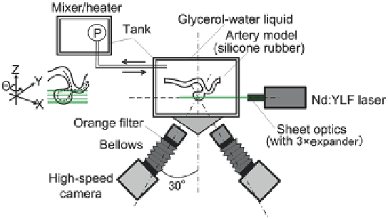
Fig. 7.28
High-magnification index matching stereo PIV setup. In-vitro experimental setup is
achieved by immersing a transparent phantom model of the aneurysm into a glycerol-water mix-
ture, which serves as the blood analogue. This setup can enable the measurement of velocity flow
field at multiple cross-sections through the aneurysm
Search WWH ::

Custom Search