Biomedical Engineering Reference
In-Depth Information
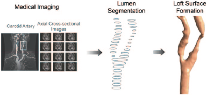
Fig. 7.1
Three-dimensional reconstruction of carotid bifurcation. Magnetic resonance imaging
was carried out on the neck region of the patient whose carotid artery can be imaged at axial orien-
tation for multiple slices. Segmentation based on the threshold of the blood vessel at various slices
is performed in the initial stage. The segmented voxels can be grouped to form a three-dimensional
anatomy and a mesh reconstruction based on the contours of these segmented regions is carried out
interest are extracted, the voxels are grouped together to form a 3D geometry. Then
the reconstruction of carotid bifurcation anatomy into a Computer Aided Design
(CAD) model is performed based on the segmentation information. The reader may
refer to the detailed procedure of medical image reconstruction from Chap. 3.
High resolution magnetic resonance imaging of a stenosed carotid bifurcation
was acquired through scanned images performed on a 42 year-old male using a
1.5-T General Electric scanner. A total of 112 contiguous slices were generated
from the high-resolution T-1 weighted spoiled gradient echo with parameters as
follows: TR, 35 ms; TE, 7 ms; flip angle, 35°; field of view, 24 cm; voxel size
0:63 mm×0:73 mm×0:63 mm. An automated detection algorithm was then ap-
plied to the images to generate a conservative segmentation of stenosed carotid
bifurcation using a two-dimensional watershed transform form markers applied to
each slice (Meyer and Beucher 1990a). Based on segmentation of different adjacent
planes, a high resolution three-dimensional computer artery model was created and
the data was stored in a stereolithography (STL) file format. To enhance the resolu-
tion of PIV measurement, the flow phantom was enlarged to 10 times of its human
counterpart. Figure
7.2
shows the reconstructed carotid geometry employed in this
case study. Based on the geometrical data from the STL file, a negative model was
created by a Rapid Prototyping (RP) printer (model ZCorp 3D) using a water-sol-
uble plaster. Following the method adopted by Hopkins et al. (2000) five layers of
water-soluble glue were painted on the plaster model to smooth and seal the pores
on its surface.
The painted prototype was then encased in a Plexiglas box where clear silicone
(using Dow Sylgard 184) was casted around the plaster model to create an optically
clear positive model. After the silicone has been cured, the plaster model was then
dissolved out with cold water, leaving a patient-specific replica of the stenosed carotid
Search WWH ::

Custom Search