Geology Reference
In-Depth Information
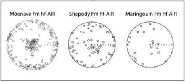
Fig. 2.3
High fi eld anisotropy of isothermal remanence (hf-AIR) for red bed formations. From left to right: the Triassic
Moenave Formation from the Colorado Plateau and the Carboniferous Shepody and Maringouin Formations from Nova Scotia.
The minimum principal axes (circles) are near to perpendicular to bedding and the maximum (squares) and intermediate axes
(triangles) are near to the bedding plane in these equal-area nets. The remanence anisotropy shows the fabric of the
ChRM-carrying hematite particles. Figures from Bilardello & Kodama (2009b) and McCall & Kodama (2010).
compared to the foreset beds of cross-bedding in red
beds, suggesting a DRM. This interpretation is based on
early laboratory re-deposition experiments that showed
the effect of initially dipping surfaces on the direction
of the DRM acquired (King 1955; Griffi ths
et al
. 1960 ).
Others have used scanning electron microscopy (SEM)
to examine the magnetic minerals in a red bed showing
multiple generations of authigenic hematite (Walker
et al
. 1981). This result strongly supports a secondary
chemical remanence for red beds. Recently, anisotropy
of magnetic remanence fabrics we have collected from
red beds show a strong depositional/compactional
fabric with minimum axes clustered perpendicular to
bedding and maximum and intermediate axes scat-
tered in the horizontal (bedding plane) (Figs 2.3 and
5.9). This fabric is very similar to that observed for
magnetite-bearing sedimentary rocks (Fig. 2.4; see
also fabric for the Perforado Formation, Fig. 5.6 and the
Pigeon Point Formation, Fig. 5.3) and strongly sup-
ports the notion that some, maybe most, red beds carry
a DRM rather than a CRM.
Can traditional DRM theory explain the remanence
of hematite-bearing rocks? The spontaneous magneti-
zation of hematite is nearly 200 times less than that of
magnetite, so is it possible that the torque of the geo-
magnetic fi eld on hematite nano-particles would be
reduced enough to preclude a prediction of perfect
alignment with the fi eld? Using reasonable values for
the spontaneous magnetization of hematite, the vis-
cosity of water and the geomagnetic fi eld results in
alignment times of 0.05 sec. This is still embarrassingly
short, and comparable to the alignment times for mag-
netite particles. In this calculation the magnetic
moment of the nominally 1.0 μm diameter hematite
particle is larger than that of the 1.0 μ m magnetite
grain in the earlier calculation because the hematite
grain is assumed to be single domain rather than
pseudo-single domain. This assumption is made
because of hematite's low spontaneous magnetization
and the importance of micro-crystalline anisotropy
controlling remanence of a hematite particle (Butler
1992 ; Tauxe 2010 ).
πη
d
mB
3
3 14159
.
××
10
−
6
10
−
3
t
=
=
=
005
.
sec
0
1 2
.
×
10
−
15
×
50
Still, red beds typically have magnetizations about an
order of magnitude stronger than magnetite-bearing
sediments (10 mA/m for red beds versus about 1 mA/m
for magnetite-bearing sediments), despite the fact that
hematite has a much lower spontaneous magnetiza-
tion than magnetite. This could suggest that hematite
grains in a sedimentary rock are better aligned than
magnetite grains.
Another way of investigating this question is to
apply the approach used by Tauxe
et al
. (2006) in their
re - deposition experiments. Tauxe
et al
. found a ratio of
DRM: SIRM for re - deposited magnetite - bearing sedi-
ments that was of the order 10-30%, where SIRM is
the saturation isothermal remanent magnetization
that is applied to the rock sample in the laboratory. It
is a measure of the total number of remanently mag-


Search WWH ::

Custom Search