Geology Reference
In-Depth Information
Neel temperatures, the temperature at which they lose
their spontaneous magnetization, to identify the mag-
netic minerals in a rock. In a standard Lowrie test,
three IRMs are applied to a rock sample in orthogonal
directions. The highest - fi eld IRM is always applied fi rst,
then the intermediate-fi eld IRM followed by the lowest-
fi eld IRM. Typical fi elds used are 1 T,
c.
0.5 T and 0.1 T,
respectively, since they are the coercivities observed for
hematite or goethite at the high end and magnetite
at the low end. The intermediate-fi eld IRM chosen
depends on the magnetic minerals suspected in the
rock or sediment. For example, pyrrhotite can some-
times have intermediate coercivities so a fi eld of 0.5
or 0.6 T can be chosen. Magnetite has a theoretical
maximum coercivity of 0.3 T for very long needle-
shaped particles, so 0.3 T can be chosen in that case.
The three-IRM rock sample is then thermally demag-
netized to observe the unblocking-temperature behav-
ior of the different IRMs in the sample. The combination
of coercivity and unblocking temperature is a powerful
identifi cation tool. For instance, a 580°C loss of mag-
netization for the 0.1 T IRM is pretty clear evidence of
magnetite, while a 680°C loss of magnetization for
the 1 T IRM is good evidence for hematite. Fe sulfi des,
usually with low to intermediate coercivities, lose their
magnetization at 300°C while goethite loses its high
coercivity IRM (> 1 T) at
c.
120 ° C.
A very powerful way of identifying the magnetic
minerals in a specimen and determining whether they
are primary depositional minerals or secondary authi-
genic minerals is by simply looking at them. Examina-
tion of the minerals
in situ
would of course be
preferable, but their concentration is so low and their
size so small (submicron) that it is not always practical
for routine analysis.
Suk
et al
. (1990) observed the secondary magnetite
in situ
from remagnetized Late Paleozoic carbonates
from Tennessee with TEM and SEM, but this was the
focus of a detailed and painstaking study of the Kiaman
remagnetization in eastern North America. For more
routine analysis as part of a standard paleomagnetic
study of sedimentary rocks, magnetic extraction of the
magnetic minerals in a sediment or rock is the best way
to characterize the main magnetic minerals.
Hounslow & Maher (1996) have outlined various
techniques in detail, but they usually involve circulat-
ing a slurry made from the disaggregated rock or from
the sediment past a strong magnetic fi eld gradient
created by small intense magnets, usually rare Earth
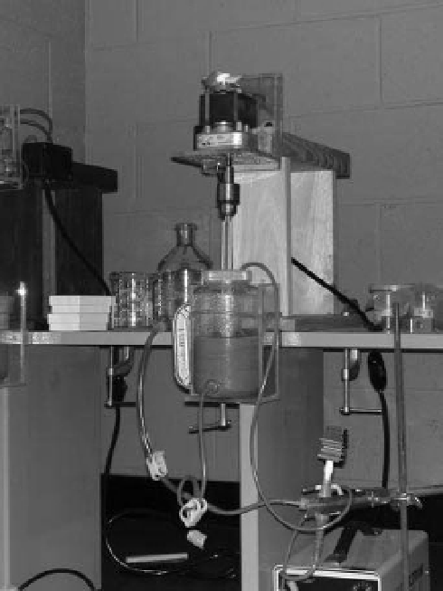
magnets (Fig. 9.3). It is often diffi cult to liberate and
Fig. 9.3
Example of a magnetic extraction set-up. Slurry
from a pulverized red bed was centrifuged to produce the
red-colored material in the device. Hematite particles were
extracted and their individual particle anisotropy was
measured (Kodama 2009). (See Colour Plate 27)
extract all the magnetic minerals in a rock and the
strongest magnetic minerals (usually magnetite) are
preferentially removed; these points must be kept in
mind when interpreting the examination of the extract
under an electron microscope.
A combination of centrifuging and magnetic extrac-
tion from a sediment slurry has been used to extract
more weakly magnetic hematite particles from a red
bed (Dekkers & Linssen 1991; Kodama 2009). The
magnetic extract can be examined under SEM or TEM.
More perfectly shaped crystals suggest authigenesis of
secondary minerals; more rounded irregularly shaped
grains suggest sedimentary transport and primary
depositional minerals. Chains of perfect single-domain
-sized magnetite or greigite crystals obviously indicate
formation by magnetotactic bacteria. The magnetic


Search WWH ::

Custom Search