Biology Reference
In-Depth Information
A
B
D
C
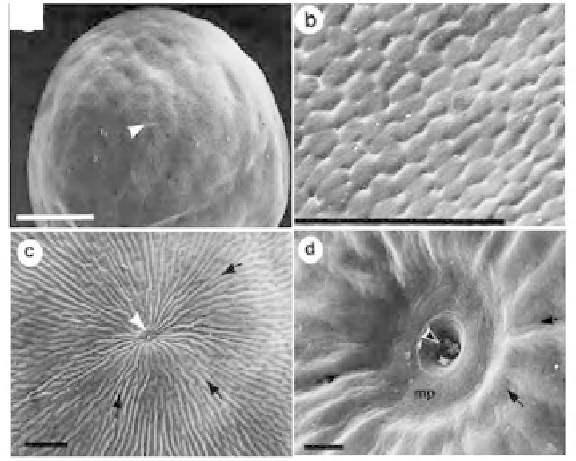
Fig. 24.
Synbranchus marmoratus
: SEM pictures of ovulated oocyte: (A) Apical view, (B)
Vegetative pole surface view, (C) Magnifi ed view of animal pole showing furrow-like
structures that converge (indicated by arrows) in the micropyle and (D) Micropylar canal
opens in the center of the micropyle pit. Scale A to C = 100 µm and d = 10 µm (from Ravaglia
and Maggese, 2002)
furrow-like structures with a slightly spiraled direction (Fig. 24C). These
furrows converge directly into micropyle pit. The micropylar canal is located
in the center of this pit (Fig. 24D) (Ravagla and Maggese 2002).
Information on morphological patterns of furrow architecture guiding
sperm to the micropylar pit is available for the eggs of many fi shes. For
instance, an incredible chorion architecture has been described with an
array of ridges and furrows in the animal pole, all of them with spiraled
arrangement and convergence into the micropyle canal of
Luciocephalus
sp.
(Riehl and Kokoscha, 1993). In the catfi sh
Sturiostoma aureum,
Riehl and
Spatzner (1991) have found eggs, whose vitelline envelope has furrows
running from vegetal to the animal pole. Using time-lapse video and image
analysis of sperm movement in the eggs of cyprinid
Barbus conchonius
,
Amanze and Iyengar (1990) have estimated that the grooves guide 99.7%
sperm into the micropyle penetration and/or fertilization.
The length and diameter of the micropylar canal differ from species to
species (e.g., Blanc et al., 1993). Yet successful heterospecifi c inseminations
occur, for instance, between
Cyprinus carpio
and
Ctenopharyngodon idella
,
suggesting the canal's diameter and length are similar in both of them.
Such reciprocal insemination is prevented in other species. For instance, the
long canal with narrow anterior opening in the eggs of Bueno Aires tetra