Biology Reference
In-Depth Information
Chapter 4
A
B
C
D
E
spermatogonia
prim spermatocytes
apoptosis
25
20
1.5
1.0
0.5
0.0
*
*
mlh1
+/+
mlh1
-/-
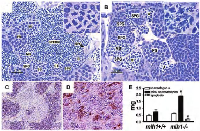
Fig. 60.
Cross sections of seminiferous tubule in (A)
mlh1
+/+
,
(B)
mlh1
-/-
zebrafi sh, showing
spermatogenic cysts with different types of germ cells, spermatogonia (SPG), primary
spermatocytes (PS), fi rst meiotic division (MI), secondary spermatocytes (SS), second meiotic
division (MII), spermatids (ST), spermatozoa (SPERM), and apoptotic spermatocytes (A). Note
the absence of postmeiosis I stages and the presence of apoptotic spermatocytes with strongly
condensed nuclei in
mlh1
-/-
testis. Inset shows a magnifi ed view of the fi rst meiotic division. In
the wild type, chromosomes are aligned at the cell equator just prior to division. In the mutant,
chromosomes are randomly distributed throughout the nucleus. TUNEL staining of (C)
mlh1
+/+
and (D)
mlh1
-/-
. High numbers of apoptotic spermatocytes are seen in mutant testis. Wild-type
testis shows a very low incidence of apoptosis. (E) Morphometric analysis of testes sections
showing increase in amounts of spermatocytes and apoptoic cells, but not of spermatogonia.
(from Feitsma et al., 2007, with permission by the Genetic Society of America)