Biomedical Engineering Reference
In-Depth Information
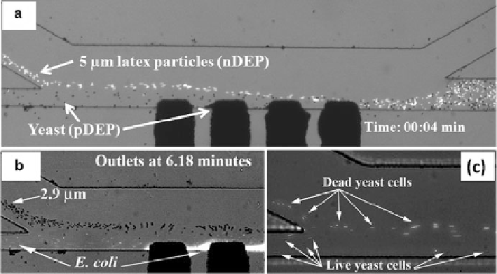
Fig. 11.5
Snapshot images of continuous separation of samples of similar sizes by polarizability
[
23
]. (
a
) Separation of yeast cells (
black dots
) by pDEP from 5
m
m fluorescent latex particles
(
white dots
) experiencing nDEP under 31.2 Vac at 300 kHz after 4 min. (
b
) Separation of stained
E. coli
cells by pDEP from 2.9
m
m latex particles exhibiting nDEP under 40.4 Vac at 1 MHz after
6.18 min. (
c
) Separation of live yeast cells by pDEP from dead yeast cells undergoing nDEP under
26.5 Vac at 1 MHz. The microscopic objective lenses used in (
a
), (
b
), and (
c
) are 5
,10
, and
20
, respectively (Reprinted with permission from Lewpiriyawong et al. (2011).
#
2011 Ameri-
can Chemical Society)
of up to 97%. In these experiments, the time window defined by the velocity of the
fluid flow (123
m
m/s) and the separation channel length (1,400
m
m) was about
11.4 s, thus resulting in an nDEP drift velocity of about 4.4
m/s.
In addition to the separation of biological cells from latex particles, the device
was also tested to separate live and dead yeast cells based on the difference in their
electrical properties. The demonstration was performed in a 600
m
S/cm NaCl
solution. Huang et al. found that the conductivity of the nucleus of live yeast
cells dramatically decreased from 0.2 to 7
m
10
3
S/m after they died [
28
]. The
reason the nucleus's conductivity rapidly changes is because the membrane of dead
yeast cells becomes more permeable and it permits content inside the cells to
exchange with the external liquid medium [
34
]. Therefore, the dead cells are less
conductive and hence are likely to experience nDEP in comparison with the live
cells. Consistent with these experimental facts, our characterization results (images
not shown here) revealed that at 1 MHz, live cells suspended in a 600
S/cm NaCl
solution underwent pDEP while dead cells underwent nDEP. Figure
11.5c
shows
continuous-flow separation of live yeast cells (black dots) from dead ones (white
dots) under 26.5 V at 1 MHz. Dead cells were repulsed from the electrodes by nDEP
and transported to the upper outlet D, while live cells under pDEP were attracted to
m
Search WWH ::

Custom Search