Biomedical Engineering Reference
In-Depth Information
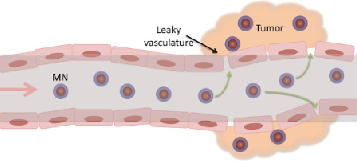
Fig. 1.2
A scheme of passive targeting with magnetic nanoparticles (MN) through the leaky
tumor vasculature
An alternate method of imaging biological phenomena with nanoparticles utilizes
the magnetic relaxation switching (MRS) property of iron oxide nanoparticles.
In this method the nanoparticle-target molecule assembly increases the ability to
change T2 relaxation time. Such a change makes these nanoparticles a better
T2 contrast agent. Consequently cancer-related biomaterials could be detected
by MRI.
1.3.1 Passive-Targeted Imaging
Even though this approach does not involve cellular specificity, it has been used
successfully in clinical trials [
24
]. Vascular permeability plays a key role in the passive
targeting strategy employed in delivering the iron oxide nanoparticles to tumor tissues.
Nanoparticles are synthesized without any targeting group attached. The nanoparticles
penetrate the tumor mass after passing through the leaky vasculature and in most cases
are retained in the tumormass. On the other hand, normal vasculature has low leakiness
and normal tissues have a tightly packed structure, which presents a barrier to
nanoparticles attempting to penetrate healthy tissues, Fig.
1.2
.
For both passive targeting and active targeting it is important to have the
nanoparticles circulate a long time in the blood stream, avoiding phagocytosis.
Macrophages eliminate foreign materials in the body, once recognized, and therefore
decrease the tumor uptake of the nanoparticles. Therefore, it is important to prevent
phagocytosis by (1) using a hydrophilic coating like PEG or dextran, (2) limiting the
size of the nanoparticles to between 30 and 50 nm, which in most cases is optimum
for passive targeting [
30
]. With such approaches it is often possible to utilize passive
targeting for cancerous tumors. Small iron oxide nanoparticles have been used for the
Search WWH ::

Custom Search