Biomedical Engineering Reference
In-Depth Information
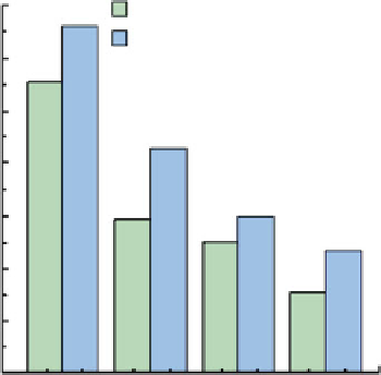
Fig. 9.18
Average thickness
of the cell-free layer at
different Hcts and diameters
[
30
,
33
]
28
PDMS micro-channel (75
μ
m)
GLASS micro-channel(100
μ
m)
24
20
16
12
8
4
0
3
2 3 5
Hct (%)
23
24
37
35
9.5 Conclusion and Future Directions
The recent developments in computing, digital image processing techniques and
microscopy have made it possible to combine both particle image velocimetry
(PIV) and particle tracking velocimetry (PTV) system with confocal microscopes,
in so-called confocal micro-PIV/PTV. In this chapter we have presented the most
relevant aspects of research using this approach: the theoretical and technical issues
for both conventional and confocal micro-PIV/PTV methods and hemodynamic
studies in micro-channels are facilitated by using systems based on these methods.
By using a 100-
m square micro-channel, the confocal micro-PIV system has
shown good accuracy in measuring blood plasma flow with Hct up to 9%. Never-
theless, for Hct higher than 9%, the light absorbed and scattered by the RBCs
contributes to a diminution of the concentration trace particles and as a conse-
quence generates spurious errors in the velocity profiles. This effect becomes much
more evident for Hct above 20%. Owing to its optical sectioning ability and
consequent improvement of the image contrast and definition, a confocal micro-
PTV system was then proposed to track individual blood cells at both low and high
Hcts. By using such a system it was indeed found possible to measure cell-cell
hydrodynamic interaction, RBC orientation and RBC radial dispersion at different
depths and Hcts. Generally, the results suggest that the RBC paths are strongly
dependent on the Hct and therefore the RBC dispersion coefficient tends to increase
with increase in Hct. In addition, the confocal micro-PTV system has proved to be a
powerful tool to obtain further insight into the flow behaviour of blood through
complex geometries such as bifurcations, confluences and stenoses. Moreover, by
culturing endothelial cells within PDMS microfluidic devices, we expect to develop
in the near future a flow system device that closely mimics the in vivo micro-vessel
environment.
m
Search WWH ::

Custom Search